
Figures & data
Figure 1. The effects of gastrodin on blood pressure and body weight in angiotensin II (Ang II)-infused mice. A tail-cuff plethysmograph method was used to measure blood pressure in mice once a week for 4 weeks, including (A) systolic blood pressure (SBP), (B) diastolic blood pressure (DBP) and (C) mean arterial pressure (MAP). (D) Body weights of mice in each group. All values are presented as mean ± standard deviation (SD). *p < 0.05 Ang II vs. control group; #p < 0.05 Ang II + gastrodin vs. Ang II group.
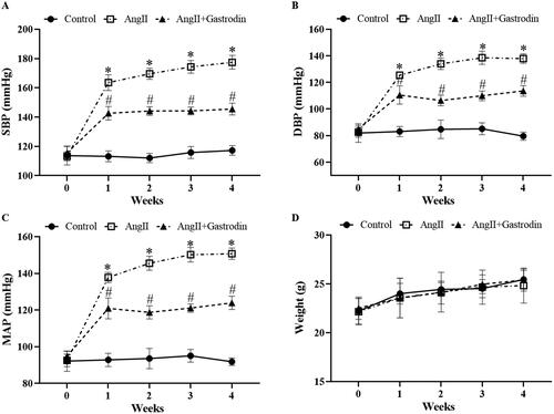
Figure 2. The effects of gastrodin on vascular function and pathological changes in the abdominal aorta of Ang II-infused mice. Ultrasound and haematoxylin and eosin (H&E) staining were performed to determine pulse wave velocity (PWV) and thickness of the abdominal aorta wall in each group. (A) PWV of the abdominal aorta. (B) Thickening of the abdominal aortic wall measured by ultrasound. (C) Representative ultrasonography images of the abdominal aorta. (D) Representative cross-sections of abdominal aortic tissues stained with H&E. Images were taken at a magnification of ×400. All values are presented as mean ± SD. *p < 0.05 Ang II vs. control group; #p < 0.05 Ang II + gastrodin vs. Ang II group.
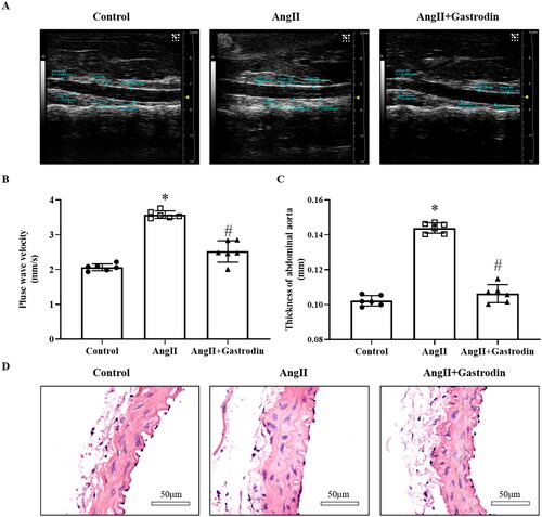
Figure 3. Gene Ontology (GO) enrichment and Kyoto Encyclopedia of Genes and Genomes (KEGG) signalling pathways analysis. (A) Hierarchical clustering plots and (B) volcano plots were used to compare gene expression profiles between the Ang II + gastrodin and Ang II groups (|fold change| ≥ 2, *p < 0.05). (C) KEGG pathway enrichment analysis of differentially expressed transcripts (DETs) comparing the Ang II + gastrodin and Ang II groups. (D–F) GO analysis was performed based on DETs from comparisons of the Ang II + gastrodin and Ang II groups. The top 30 enriched items for (D) biological processes, (E) cellular composition and (F) molecular function are presented.
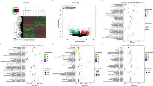
Figure 4. The effects of gastrodin on contraction and relaxation of the abdominal aorta. An Automated Multi Myograph System was performed to measure vasorelaxation of abdominal aortic rings. (A) Vascular contraction at baseline after gastrodin treatment. (B) The effect of gastrodin on Ang II-induced contraction in abdominal aortic rings. (C) The effect of gastrodin on relaxation of abdominal aortic rings precontracted with norepinephrine (NE). (D) The effect of verapamil on gastrodin-induced vasorelaxation. All values are represented as mean ± SD; *p < 0.05 vs. control group.
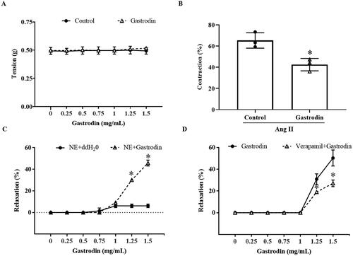
Figure 5. Expression of proteins involved in the MLCK/p-MLC2 pathway in abdominal aorta tissues. Immunohistochemical (IHC) staining was used to detect protein levels of (A) phospho-myosin light chain 2 (p-MLC2), (B) myosin light chain 2 (MLC2), (C) angiotensin type 1 receptor (AT1R), (D) calmodulin (CaM) and (E) myosin light chain kinase (MLCK) in abdominal aorta tissues. Images were taken at a magnification of ×400. All values are represented as mean ± SD; *p < 0.05 Ang II vs. control group; #p < 0.05 Ang II + gastrodin vs. Ang II group.
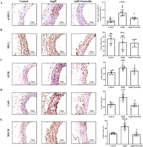
Figure 6. The effects of gastrodin on release of intracellular calcium in Ang II-stimulated vascular smooth muscle cells (VSMCs). Confocal microscopy was performed to measure changes in intracellular Ca2+ concentration in real time. (A) Representative images of intracellular calcium release in VSMCs at different time points. Images were taken at a magnification of ×200. (B) A graph of the change in intracellular Ca2+ concentration is shown.
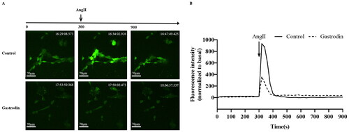
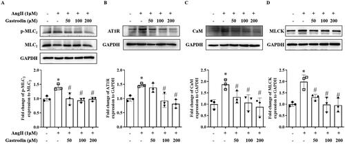
Figure 7. The effects of gastrodin on the MLCK/p-MLC2 pathway in Ang II-stimulated VSMCs. Western blotting was used to determine protein levels of (A) p-MLC2, (B) AT1R, (C) CaM and (D) MLCK in Ang II-stimulated VSMCs pretreated with gastrodin (50, 100 or 200 μM) for 24 h. All values are represented as mean ± SD; *p < 0.05 Ang II vs. control group; #p < 0.05 vs. Ang II group.
Supplemental Material
Download MS Word (813.3 KB)Data availability statement
The datasets used and/or analysed during the current study are available from the corresponding author on reasonable request.