Figures & data
Figure 1. Protocols for the papillary muscle experiments. BIM: bisindolylmaleimide 20 nM, BK: bradykinin 500 nM, ISO: isoproterenol 100 nM, WM: wortmannin 100 nM ↓: 3 min exposure to ISO.
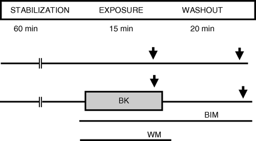
Figure 2. Contractile force amplitude in response to isoproterenol in papillary muscle treated with bradykinin in combination with inhibitors to PKC or PI3-kinase. BIM: bisindolylmaleimide 20 nM, BK: bradykinin 500 nM, ISO: isoproterenol 100 nM, WM: wortmannin 100 nM: p < 0.05 compared to control.
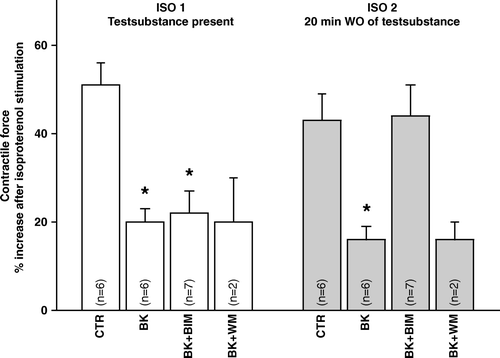
Table I. Contractile force amplitude. Initial values.
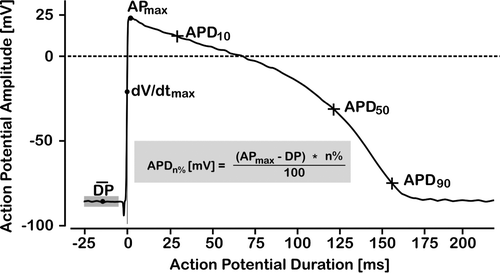
Figure 3. Simulated guinea pig action papillary muscle potential illustrating how APD-10, APD-50 and APD-90 were determined. Time 0 was set at maximum rate of rise of phase 0 depolarization, baseline resting membrane potential was set as the average of a 20 ms sampling sequence at 50 kHz starting at time −25 ms.