Figures & data
Figure 1. Rabbits, chloralose anesthesia. Series 1. Pre-occlusion levels of heart rate, mean arterial pressure and s.d. of R-R intervals per respiratory cycle.
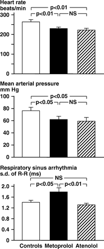
Table I. Rabbits anesthetized with chloralose – Series 1.
Table II. Rabbits anesthetized with chloralose – Series 1.
Figure 2. Rabbits, chloralose anesthesia. Series 1. Rabbits (percent) surviving in the three treatment groups during the 30-minute period after coronary artery occlusion. The difference in survival between the metoprolol animals and the other two groups was statistically significant (p < 0.05).
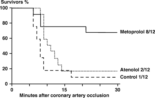
Figure 3. Rabbits, chloralose anesthesia. Series 1. Pre-occlusion values for heart rate and s.d. R-R per respiration in controls (Δ), metoprolol (○) and atenolol rabbits (□). Filled symbols: Animals, which fibrillated after the subsequent coronary artery occlusion. Open symbols: Animals without VF after the coronary artery occlusion.
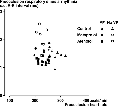
Table III. Rabbits anesthetized with chloralose – Series 2.