Figures & data
Table I. Demographics and clinical data of the patients treated with Placebo, VEGF, low dose G-CSF (lG-CSF) and VEGF-A165- combined with high dose G-CSF (hG-CSF).
Figure 1. A) Circulating CD34+ cells (103/mL) from baseline to day 28. B) Increase in circulating CD45 − CD34− relative to the baseline value in each group. Data in panel A are previously published. Citation12, Citation13
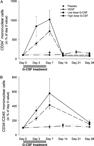
Figure 2. Relative increase in circulating CD45 − CD34− subtype (A) CD105+ and CD45-CD34- cell types with endothelial markers (B) CD31+ (C) CD144+ (D) KDR+ and (E) CXCR4+ in the first month after G-CSF treatment. *p < 0.05 day 3 and day 7 versus day 1 in low dose group. #p < 0.05 day 3 and day 7 versus day 1 in high dose group.
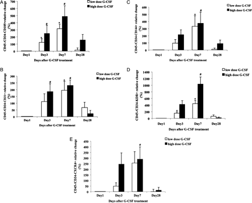
Table II. Plasma concentrations of VEGF and stromal-derived factor-1 (SDF-1) in patients treated with placebo, plasmid VEGF-A165, G-CSF or VEGF-A165- combined with G-CSF the first month after treatment.
Table III. Correlations between peak value of mobilized cells and change in clinical characteristics from baseline to follow-up for patients treated with G-CSF or VEGF-A165- combined with G-CSF.