Figures & data
Figure 1. Part A) is a schematic illustration of LV emptying (outflow), and filling (inflow) in a LV volume versus time plot. Part B) is achieved by differentiation of the plot in A). The first derivative; flow (volume/time) is plotted against time for assessment of the velocities of emptying and filling and the corresponding acceleration of these velocities. In- and outflow were found to be the most appropriate terms since flow equals volume per time unit. Explanation: 1) Maximum outflow acceleration; 2) Maximum outflow; 3) Time to maximum outflow; 4) Time to end-systole; 5) Maximum early inflow acceleration; 6) Maximum early inflow; 7) Time to maximum inflow; 8) Maximum late inflow; 9) Time to maximum late inflow.
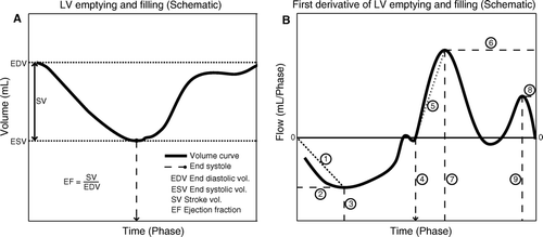
Table I. Global CMR parameters, and systolic- and diastolic-derived functional parameters
Figure 2. CMR images of LV long-axis in end-diastole (left column) and end-systole (right column) of an intervention animal (top row) versus a control animal (bottom row). The infarction is marked by a dashed line and arrows and is clearly seen to involve the septum, the apex, and the free wall of the LV.
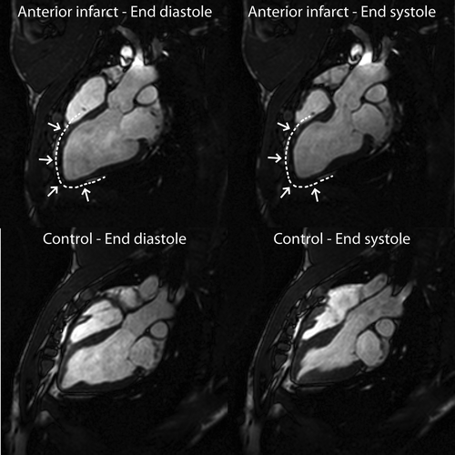
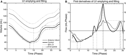
Figure 3. Part A) shows LV volume versus time plot for intervention animals (grey line) and control animals (black line). Part B) shows the first derivative, flow versus time with the same color coding. See text for description of results. In both A) and B) the time signature is cardiac phase; this is done to avoid bias due to differences in heart rate amongst groups as described in the text.