Figures & data
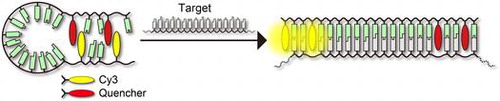
Figure 1. Sequences of oligonucleotides used in this study. Chemical structures of dyes and D-threoninol linker used to incorporate dyes into oligonucleotides are shown.
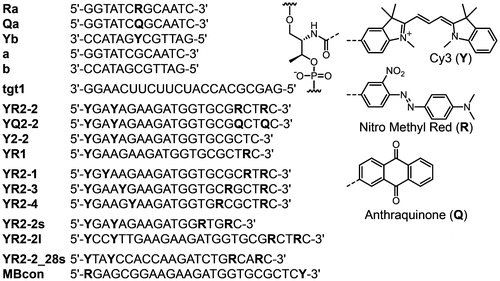
Figure 2. (A) Fluorescence spectra of model duplexes a/Yb, Qa/Yb, and Ra/Yb. Conditions: 2.0 μM quencher strands and 1.0 μM Yb in 100 mM NaCl, 10 mM phosphate buffer (pH 7.0), 20 °C. (B) UV-visible absorption spectra of single-strands Ra and Yb, and Ra/Yb duplex. Summation of spectra of Ra and Yb is shown in blue dotted line. Conditions: 2.0 μM each strand, 100 mM NaCl, 10 mM phosphate buffer (pH 7.0), 20 °C.
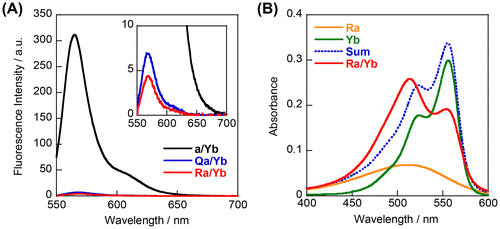
Figure 3. Emission spectra (A) without and (B) with RNA target of probes YR2–2, YQ2–2, YR1, and Y2–2. Conditions: 0.2 μM probe, 0.4 μM tgt1, 100 mM NaCl, 10 mM phosphate buffer (pH 7.0), 20 °C.
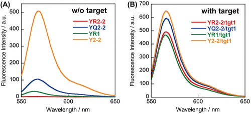
Figure 4. Effects of temperature on UV-visible absorption spectra of YR2–2 (A) without or (B) with target RNA. Conditions: 1.0 μM each strand, 100 mM NaCl, 10 mM phosphate buffer (pH 7.0).
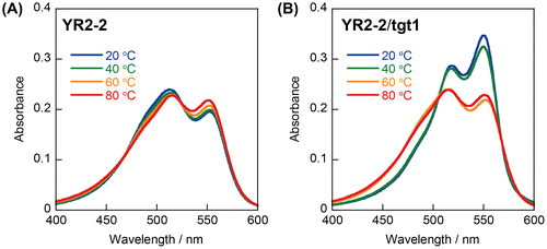
Figure 5. Responses of YR2–2 and conventional molecular beacon (MBcon) to the target (tgt1). Target RNA was added at the point of 0 s. Conditions: 0.2 μM probe strands, 0.4 μM tgt1, 100 mM NaCl, 10 mM phosphate buffer (pH 7.0), 20 °C.
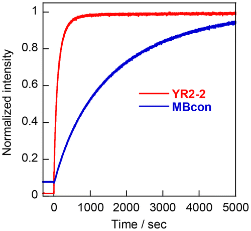
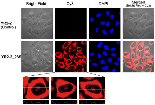
Figure 6. Confocal microscopy images of fixed and permeabilized HeLa cells treated with YR2–2 or YR2–2_28S without washing prior to imaging. Nuclei were stained with DAPI. Intensity analyses of cells are shown at the bottom.
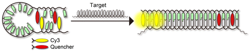
Scheme 1. Schematic illustration of our strategy to detect RNA by using a stem-less probe.