Figures & data
Figure 1. TEM images (scale bar = 100 nm) and magnified section (scale bar = 5 nm) with corresponding SAED patterns (scale bar = 5 1/nm) of (a, e) PHAp; (b, f) 0.25-FPHAp; (c, g) 0.50-FPHAp; (d, h) 0.75-FPHAp. SEM images (scale bar = 1 μm) of (i) PHAp; (j) 0.25-FPHAp; (k) 0.50-FPHAp; (l) 0.75-FPHAp. EDS spectra, as well as the distribution of calcium and fluoride by EDS mapping of (m) PHAp; (n) 0.25-FPHAp; (o) 0.50-FPHAp; (p) 0.75-FPHAp.
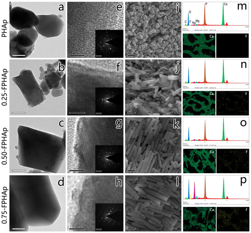
Table 1. Elemental analysis of PHAp and FPHAp samples by EDS.
Figure 2. (a) FTIR spectra, (b) expanded water stretching region and (c) OH libration region of PHAp, 0.25-FPHAp, 0.50-FPHAp, and 0.75-FPHAp. (d) XRD patterns and (e) expanded X-ray reflections of PHAp, 0.25-FPHAp, 0.50-FPHAp, and 0.75-FPHAp (all spectra are vertically offset for clarity).
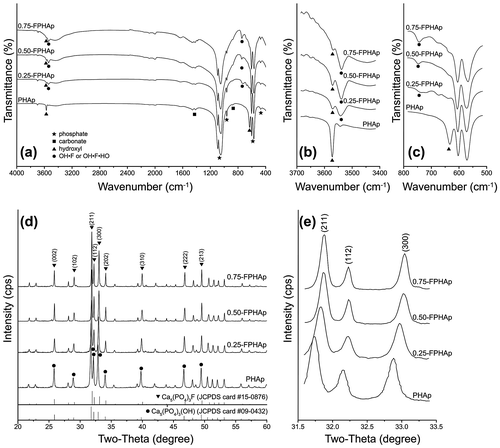
Table 2. Lattice constants and crystallinity of PHAp, 0.25-FPHAp, 0.50-FPHAp, and 0.75-FPHAp. With the rise in fluoride content, the crystallinity of the apatite steadily increased. The incorporation of fluoride also resulted in significant decline of a lattice constant of the apatite, while the c lattice constant did not show any discernable difference.
Figure 3. Box plot showing the compressive strength of PHAp, 0.25-FPHAp, 0.50-FPHAp, and 0.75-FPHAp.
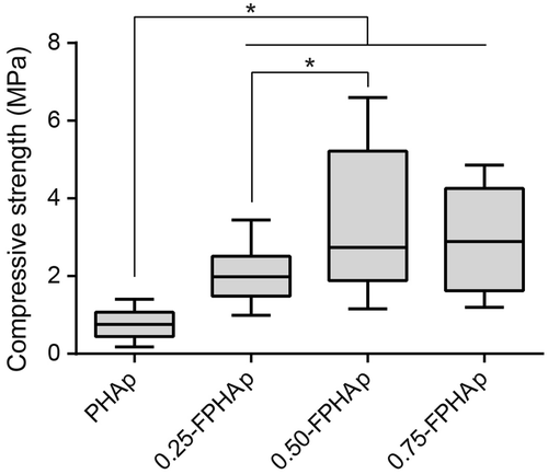
Figure 4. (a) Mercury intrusion cumulative curves of PHAp, 0.25-FPHAp, 0.50-FPHAp, and 0.75-FPHAp. (b) Incremental intrusion in relate to pore diameter showing the pore diameter distribution of PHAp, 0.25-FPHAp, 0.50-FPHAp, and 0.75-FPHAp.
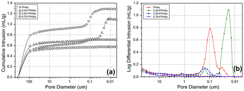
Figure 5. Solubility isotherms of PHAp, 0.25-FPHAp, 0.50-FPHAp, and 0.75-FPHAp 100 mM KCl solution at 37.0 ± 0.1°C by solid titration. Solubility data of stoichiometric HAp were shown for comparison.
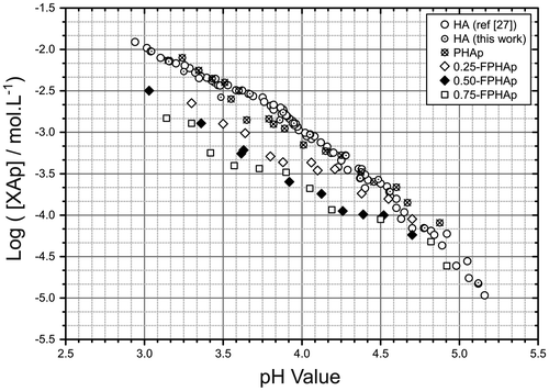
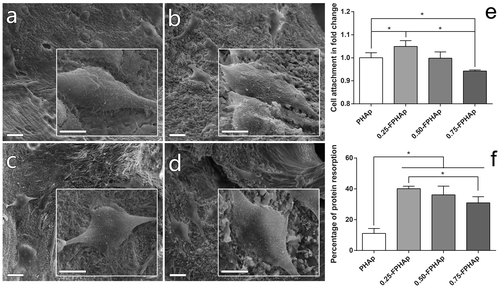
Figure 6. SEM images (scale bar = 20 μm) and inserted magnified view (scale bar = 10 μm) showing the adhesion of human osteoblastic-like MG63 cell on the surfaces of (a) PHAp, (b) 0.25-FPHAp, (c) 0.50-FPHAp, and (d) 0.75-FPHAp. (e) Quantitative evaluation of cell attachment showed only 0.25-FPHAp favored increased attachment of MG63, while 0.75-FPHAp was inferior than PHAp for cell attachment. (f) Protein absorption assay demonstrated that the incorporation of fluoride resulted in significant higher protein absorption of the apatite.