Figures & data
Figure 1. SEM images of porous chitosan scaffolds prepared at different freezing temperatures and times. Scale bar = 100 μm.
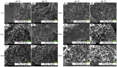
Figure 2. Pore sizes of the prepared porous chitosan scaffolds at different temperatures and times.
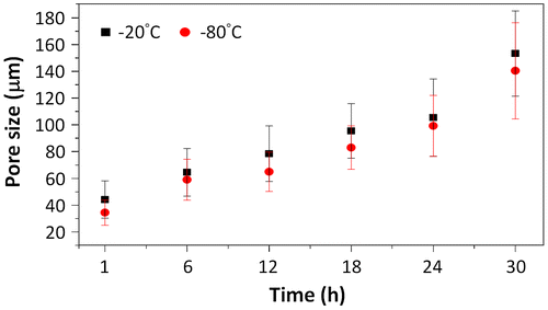
Figure 3. Viability of cells under different bFGF concentrations after seven days of culture; (a) L929 mouse fibroblasts and (b) Bovine endothelial cells. Viability in both cell types is shown relative to that of cells cultured on TCPS. Different letters indicate a significant difference when compared to the concentration labeled by that letter (p < 0.05).
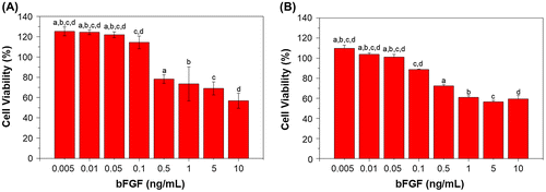
Figure 4. Viability of (a) L929 and (b) BEC cultured under different TGFβ1 concentrations for seven days. Results are shown in percent viability relative to TCPS. Letter a in (a) indicates a significant difference (p < 0.05).
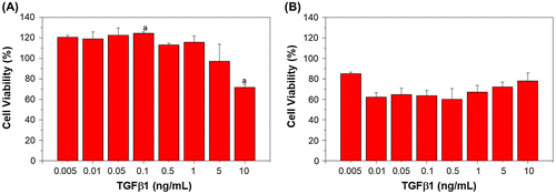
Figure 5. The cumulative release of various bFGF contents from porous chitosan scaffolds within (a) 24 h and (b) up to 21 days (1, 4, 8, 12, 24 h, and 3, 7, 14, 21 days).
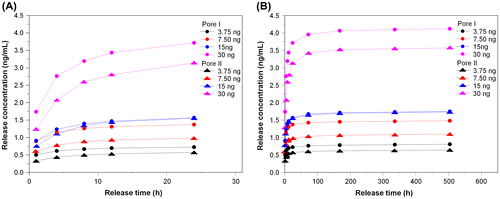
Figure 6. Single-point release of different, initial loading amounts of bFGF at different times following immersion in a solvent for (a) Pore I and (b) Pore II chitosan scaffolds. Inset: reduction trends for different loadings.
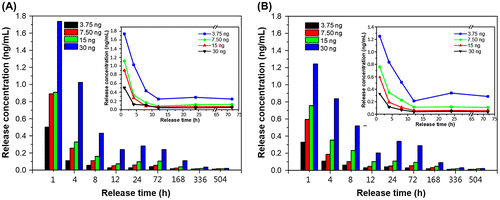
Table 1. Release characteristics of various bFGF contents from porous chitosan scaffolds.
Figure 7. The cumulative release of various TGFβ1 contents from porous chitosan scaffolds within (a) 24 h and (b) up to 21 days (1, 4, 8, 12, 24 h, and 3, 7, 14, 21 days)
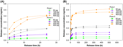
Figure 8. Single-point release of different TGFβ1 content at 1, 4, 8, 12, 24, 72, 168, 336, and 504 h for (a) Pore I and (b) Pore II chitosan scaffolds. Inset: reduction trends for different loadings.
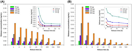
Table 2. Release characteristics of various TGFβ1 contents from porous chitosan scaffolds.
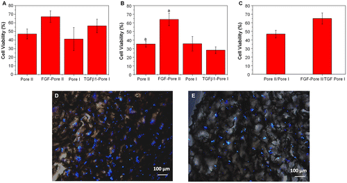
Figure 9. Effect of porous chitosan scaffolds on the proliferation of (a) L929 and (b) BEC in the presence and absence of growth factors (bFGF and TGFβ1) relative to TCPS. Letter a in (a) indicates a significant difference; (c) Cell viability of L929 and BEC co-culture in Pore I and Pore II scaffolds containing TGFβ1 and bFGF, respectively, relative to TCPS. (p < 0.05). Fluorescent images of (d) L929 and (e) BEC cultured on porous chitosan scaffolds containing growth factors bFGF and TGFβ1. Blue represents nucleus on the chitosan scaffold.