
Figures & data
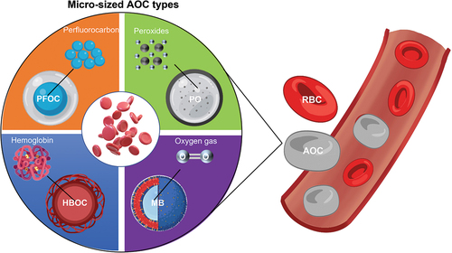
Figure 1. Human red blood cells (hRbcs).
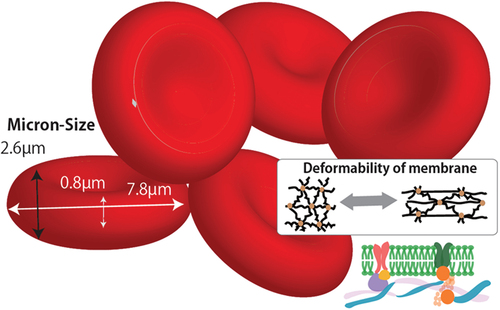
Figure 2. Micro-sized AOCs inspired by hRbcs.
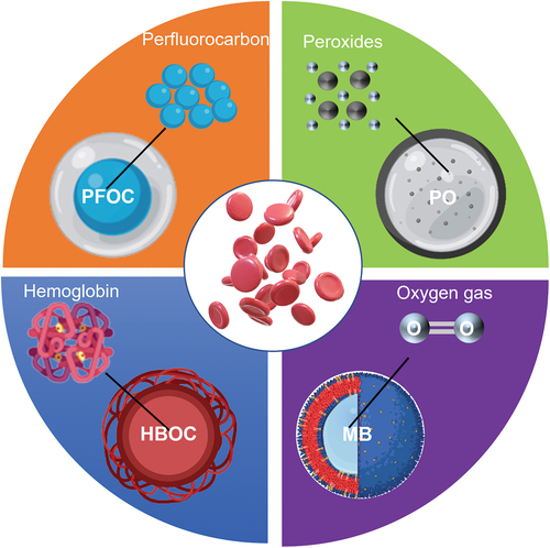
Figure 3. Typical abnormal RBC morphologies observed in the peripheral blood samples from COVID − 19 patients, adapted with permission from [Citation33]. Copyright 2022 Marchi, Bozzini, Bertolone, Dima, Busti, Castagna, Stranieri, Fratta Pasini, Friso, Lippi, Girelli and Vianello.
![Figure 3. Typical abnormal RBC morphologies observed in the peripheral blood samples from COVID − 19 patients, adapted with permission from [Citation33]. Copyright 2022 Marchi, Bozzini, Bertolone, Dima, Busti, Castagna, Stranieri, Fratta Pasini, Friso, Lippi, Girelli and Vianello.](/cms/asset/4f8ee40e-4c7b-44f0-86c8-c97c9c074ec7/tsta_a_2223050_f0003_oc.jpg)
Figure 4. Fabrication of AOCs by coprecipitation and subsequent chemical cross-linking. (a) Fabrication scheme of hemoglobin microparticles, reprinted with permission from [Citation58]. Copyright 2012, American Chemical Society (b) Fabrication scheme of Hb particles, reprinted with permission from [Citation59]. Copyright 2013, American Chemical Society (c) Schematic representation of the assembled Hb microspheres with the surface modified by PEG, reprinted with permission from [Citation60]. Copyright 2012, American Chemical Society.
![Figure 4. Fabrication of AOCs by coprecipitation and subsequent chemical cross-linking. (a) Fabrication scheme of hemoglobin microparticles, reprinted with permission from [Citation58]. Copyright 2012, American Chemical Society (b) Fabrication scheme of Hb particles, reprinted with permission from [Citation59]. Copyright 2013, American Chemical Society (c) Schematic representation of the assembled Hb microspheres with the surface modified by PEG, reprinted with permission from [Citation60]. Copyright 2012, American Chemical Society.](/cms/asset/f94d6e5d-92c4-4a92-abe9-b2b10b0506e4/tsta_a_2223050_f0004_oc.jpg)
Figure 5. Preparation of AOCs by SPG membrane emulsification. (a) Hb/BSA microspheres reprinted with permission from [Citation79]. Copyright 2015 Elsevier B.V. (b) PLC shell/PFOB core microparticles reprinted with permission from [Citation73]. Copyright 2019 American Chemical Society.
![Figure 5. Preparation of AOCs by SPG membrane emulsification. (a) Hb/BSA microspheres reprinted with permission from [Citation79]. Copyright 2015 Elsevier B.V. (b) PLC shell/PFOB core microparticles reprinted with permission from [Citation73]. Copyright 2019 American Chemical Society.](/cms/asset/7df1fecf-4274-42a4-a201-21c453d57373/tsta_a_2223050_f0005_oc.jpg)
Figure 6. Fabrication process of RBC-mimicking micro-sized AOCs from PS template by LbL method, adapted with permission from [Citation94]. Copyright 2009 National Academy of Science.
![Figure 6. Fabrication process of RBC-mimicking micro-sized AOCs from PS template by LbL method, adapted with permission from [Citation94]. Copyright 2009 National Academy of Science.](/cms/asset/b620936f-a2ec-4eea-bc9e-37855094cd93/tsta_a_2223050_f0006_oc.jpg)
Table 1. Micro-sized AOCs which were inspired by hRBC.
Figure 7. Culture systems using PFC-based AOCs. (a) Diagram of perfusion loop of oxygen supply with AOCs for culturing cardiac fibroblasts, adapted with permission from [Citation136]. Copyright 2006 UPV/EHU Press (b) Diagram of PerfusionPal system operation in which dense blood substitute acts as a piston to drive the flow of medium up and down, reprinted with permission from [Citation135]. Copyright 2020 Shoemaker et al. (c) Diagram outlining the perfluorocarbon (PFC) membrane AirHive cell culture dish compared to a transwell dish, reprinted with permission from [Citation134]. Copyright 2020 Mirza Muhammad Fahd Qadir et al.
![Figure 7. Culture systems using PFC-based AOCs. (a) Diagram of perfusion loop of oxygen supply with AOCs for culturing cardiac fibroblasts, adapted with permission from [Citation136]. Copyright 2006 UPV/EHU Press (b) Diagram of PerfusionPal system operation in which dense blood substitute acts as a piston to drive the flow of medium up and down, reprinted with permission from [Citation135]. Copyright 2020 Shoemaker et al. (c) Diagram outlining the perfluorocarbon (PFC) membrane AirHive cell culture dish compared to a transwell dish, reprinted with permission from [Citation134]. Copyright 2020 Mirza Muhammad Fahd Qadir et al.](/cms/asset/4ff40f80-668d-46a9-97af-6d87be906802/tsta_a_2223050_f0007_oc.jpg)
Figure 8. Oxygen releasing scaffold using micro-sized AOCs for new wound dressing, adapted with permission from [Citation122]. Copyright 2018 Jeongyeon Choi et al.
![Figure 8. Oxygen releasing scaffold using micro-sized AOCs for new wound dressing, adapted with permission from [Citation122]. Copyright 2018 Jeongyeon Choi et al.](/cms/asset/b63ab7dd-900c-41ea-b296-b0d659f6089f/tsta_a_2223050_f0008_oc.jpg)
Figure 9. Application of HBOC in solid organ preservation. (a) Pre-oxygenation process of HBOC-added preservation solution. (b) Static cold preservation of isolated liver after pre-oxygenation with the HBOC-added preservation solution. (c) Mechanical perfusion of the isolated liver after pre-oxygenation. Adapted with permission from [Citation14]. Copyright 2021 Cao et al.
![Figure 9. Application of HBOC in solid organ preservation. (a) Pre-oxygenation process of HBOC-added preservation solution. (b) Static cold preservation of isolated liver after pre-oxygenation with the HBOC-added preservation solution. (c) Mechanical perfusion of the isolated liver after pre-oxygenation. Adapted with permission from [Citation14]. Copyright 2021 Cao et al.](/cms/asset/7a09bf35-8e1b-4e79-92ad-bbea2128add3/tsta_a_2223050_f0009_oc.jpg)
Figure 10. Functionalization process and confocal microscopic images of Ru(ddp)-loaded FDC emulsions stabilized with 2% F127 under hypoxic condition (a,b) and normoxic condition (c,d). Scale bars: 20 μm, reprinted with permission from [Citation73]. Copyright 2019, American Chemical Society.
![Figure 10. Functionalization process and confocal microscopic images of Ru(ddp)-loaded FDC emulsions stabilized with 2% F127 under hypoxic condition (a,b) and normoxic condition (c,d). Scale bars: 20 μm, reprinted with permission from [Citation73]. Copyright 2019, American Chemical Society.](/cms/asset/0f3aedcc-874d-4dea-b435-6d924843c380/tsta_a_2223050_f0010_oc.jpg)