
Figures & data
Figure 1. PD-L1 expression in invasive high-grade urothelial carcinoma. Strong and diffuse membranous expression is seen in the epithelial cells (Right) whereas it is mostly cytoplasmic in the tumor-infiltrating immune cells (left) (PD-L1 IHC 22C3 pharmDx by Dako/Agilent).
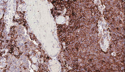
Table 1. Diagnostic PD-L1 expression assay evaluation for bladder cancer.
Figure 2. Complex interplay of chemokines and cytokines classify the inflammatory state of the tumor microenvironment. Interferon-g (IFN-g) released by activated T cells and NK cells activates STAT1, IDO-1 (indolamine oxygenase I) and CMKLR1 in dendritic cells and macrophages (1). STAT-1 mediated signaling and additional pathways produce the chemokines CCL5 and CXCL9 (2). This recruits additional T cells into the tumor microenviroment through CXCR6. IFN-g stimulates the expression of HLA molecules and proteasome components including PSMB10 (3). Finally, IFN-g upregulates a number of immune checkpoint molecules including PD-L1, PD-L2, TIGIT, LAG-3, and B7-H3 on T cells (4). Reproduced under the terms of the Creative Commons Attribution 4.0 International License (http://creativecommons.org/licenses/by/4.0/) – Figure 4a Ref [Citation18]. J Immunother Cancer. 2017; 5:94.
![Figure 2. Complex interplay of chemokines and cytokines classify the inflammatory state of the tumor microenvironment. Interferon-g (IFN-g) released by activated T cells and NK cells activates STAT1, IDO-1 (indolamine oxygenase I) and CMKLR1 in dendritic cells and macrophages (1). STAT-1 mediated signaling and additional pathways produce the chemokines CCL5 and CXCL9 (2). This recruits additional T cells into the tumor microenviroment through CXCR6. IFN-g stimulates the expression of HLA molecules and proteasome components including PSMB10 (3). Finally, IFN-g upregulates a number of immune checkpoint molecules including PD-L1, PD-L2, TIGIT, LAG-3, and B7-H3 on T cells (4). Reproduced under the terms of the Creative Commons Attribution 4.0 International License (http://creativecommons.org/licenses/by/4.0/) – Figure 4a Ref [Citation18]. J Immunother Cancer. 2017; 5:94.](/cms/asset/97656089-481c-42a5-acef-31dc8c8ad15b/iebt_a_1733965_f0002_oc.jpg)