
Figures & data
Figure 1. Reaction schemes for the making of the anti-cMET Fab drug conjugates (FDC): (1) lysine-based conjugation approach and (2) cysteine-based conjugation approach (C-lock) after a mild selective reduction of the LC/HC disulfide bond.
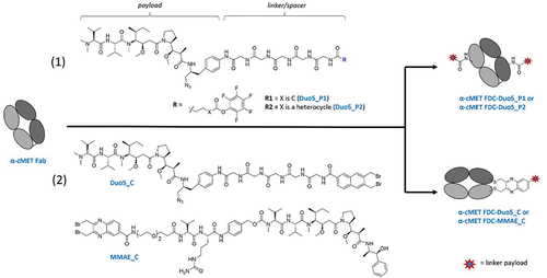
Figure 2. Characterization of the anti-cMET Fab drug conjugates. a) SDS-PAGE analyses of the lysine-based conjugation approach (left) and cysteine-based conjugation approach (right), b) binding affinity determination via ELISA of the anti-cMET Fab/IgG and their corresponding drug conjugates, c) time-course internalization study of the anti-cMET Fab in live MDA-MB-468 cells (moderate cMET expression) via confocal microscopy.
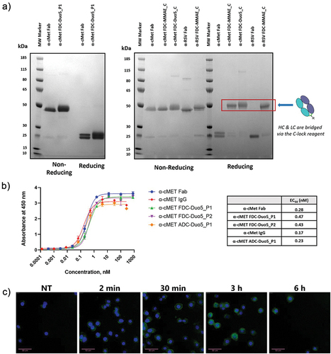
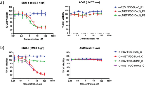
Figure 3. In vitro potency of the FDCs toward cMET high and cMET low expressing cancer cell lines. a) anti-cMET FDCs from lysine-based approach vs a non-targeting FDC (α-RSV, respiratory syncytial virus) on SNU-5 (gastric) and A549 (lung) cells, b) anti-cMET FDCs from the C-lock approach with either Duo5 or MMAE as payload on the same cell lines c) anti-cMET FDCs potency toward HT-29 (colorectal) and SK-BR-3 (breast) cancer cells d) FDC vs ADC with varying DAR.
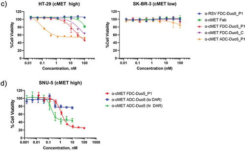
Table 1. Composition of the different FDCs/ADCs and their IC50 values.
Figure 4. In vitro characterization of induction of cMET receptor dimerization and subsequent activation of downstream effector. a) Western blot analysis of cell extracts after an overnight serum starved-A549 cells (cMET low) were stimulated with either anti-cMET Fab or anti-cMET IgG, b) cytotoxicity assay of FDC in the presence or absence of anti-cMET IgG in SNU-5 cells.
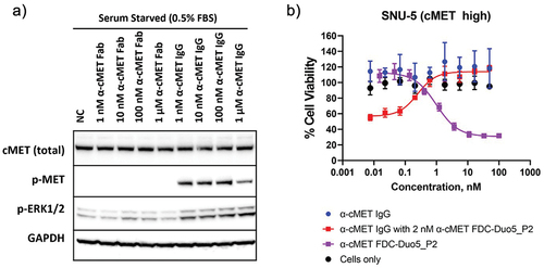
Figure 5. In vivo efficacy evaluation of the anti-cMET FDC on SNU-5 subcutaneous xenograft in Nu/Nu mice. Treatment was initiated when the average tumor size reached 150–200 mm3. Tumor size (a) and body weights (b) of mice were measured twice weekly. α-cMET FDC-Duo5_P1 significantly inhibited SNU-5 tumor growth in nude mice in a dose-dependent manner up to 88 days after initial treatments. **** indicate P value < 0.0001, two-way Anova with Tukey’s multiple comparisons test to vehicle control PBS. Data = mean ± SEM (N = 8); ns = not significant.
