
Figures & data
Figure 1. BTK signal transduction pathways. After BCR is activated by phosphorylation, it recruits spleen tyrosine kinase (SYK) to the membrane where it is phosphorylated and subsequently phosphorylates Bruton’s tyrosine kinase (BTK). Phosphorylated BTK is recruited to the plasma membrane via autophosphorylation of Tyr223 in the SRC homology domain, and phosphorylates PLCγ2. Activated PLCγ2 hydrolyzes phosphatidyl inositol 4,5-biphosphate (PIP2), which results in the generation of inositol triphosphate (IP3) and diacylglycerol (DAG). IP3 upregulates calcium levels and DAG mediates activation of protein kinase Cβ (PKCβ). Increased of calcium levels, activation of PLCγ2, and PKCβ in turn promote the activation of the NF-κB-, ERK-, and NFAT-dependent pathways, which control the transcriptional expression of genes involved in proliferation, survival and cytokines secretion. In order to facilitate the understanding of the figure, some intermediate pathways have not been drawn. Ag: antigen; BCAP: B cell adaptor for PI3K; BCR: B cell receptor; BLNK: B cell linker; BTK: Bruton’s tyrosine kinase; DAG: diacylglycerol; ERK: extracellular-signal-regulated kinase; Igα/Igβ: signal transduction moiety (CD79); IKK: IκB kinase; IP3: inositol 1,4,5-triphosphate; NFAT: nuclear factor of activated T cells; NF-κB: nuclear factor κB; PKCβ: protein kinase Cβ; PLCγ2: phospholipase C-γ2. SYK: spleen tyrosine kina
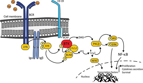
Table 1. Bruton’s Tyrosine Kinase inhibitor emerging drugs for the MS treatment