Figures & data
Figure 1. Immunolocalisation of ERα and PR in full thickness sections of human endometrium.
Images shown from four samples of uterine tissue recovered during the early (ES) or mid (MS) secretory phases of the cycle: each section represents the full thickness of the uterine wall with the lumen at the top and myometrium at the bottom. Sections were co-stained for ERα (red) and PR (green) using standard protocols [Citation32] for clarity the images recorded in the different channels (red, green) are shown side-by-side rather than overlaid. Note that during the ES there is intense immunopositive staining for both ERα and PR-A in the glandular epithelium in both the functional (arrows) and basal (white asterisks) layers. During the MS immunoexpression in the epithelium is down-regulated but expression of PR in stromal fibroblasts is maintained (green asterisks). Full color available online.
![Figure 1. Immunolocalisation of ERα and PR in full thickness sections of human endometrium.Images shown from four samples of uterine tissue recovered during the early (ES) or mid (MS) secretory phases of the cycle: each section represents the full thickness of the uterine wall with the lumen at the top and myometrium at the bottom. Sections were co-stained for ERα (red) and PR (green) using standard protocols [Citation32] for clarity the images recorded in the different channels (red, green) are shown side-by-side rather than overlaid. Note that during the ES there is intense immunopositive staining for both ERα and PR-A in the glandular epithelium in both the functional (arrows) and basal (white asterisks) layers. During the MS immunoexpression in the epithelium is down-regulated but expression of PR in stromal fibroblasts is maintained (green asterisks). Full color available online.](/cms/asset/a1ca4e90-6012-4bcb-86bc-87e8f2696491/iett_a_1180368_f0001_oc.jpg)
Table 1. Non-genomic signaling pathways reported to be triggered by progesterone.
Figure 2. Structure of common SPRMs.
Chemical structures of selective progesterone receptor modulators (SPRMs) in current clinical use or which have been in clinical development.
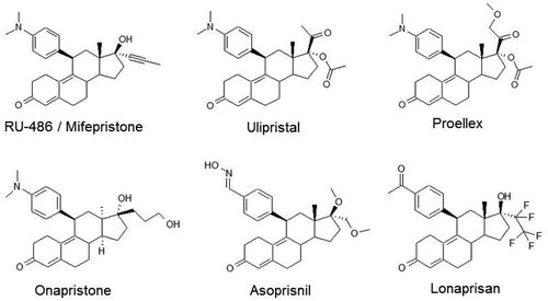
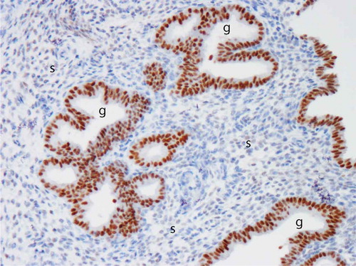
Figure 3. Image of progesterone receptor (PR) immuno-reactivity in human endometrium after administration of a selective PR modulator (SPRM). Note intense positive (brown) immunostaining in the glandular epithelium (g) and virtual absence of immuno-reactivity in the stroma (s). Image kindly provided by Professor Alistair Williams, University of Edinburgh. Full color available online.