3,653
Views
35
CrossRef citations to date
0
Altmetric
Review

Deep brain stimulation for the treatment of uncommon tremor syndromes

&
Pages 983-997 | Received 24 Feb 2016, Accepted 19 May 2016, Published online: 09 Jun 2016

Figures & data

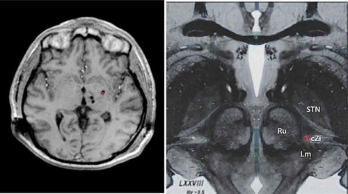
Figure 1. LEFT. T1 Weighted Axial Magnetic Resonance image showing multi-target DBS with bilateral thalamic (VIM and VoP) leads along with ipsilateral Gpi DBS (red dot). RIGHT. Upper midbrain anatomical axial image showing the relevant Posterior Subthalamic area DBS anatomy and neurosurgical target (Red X). Red Nucleus (Ru), Subthalamic nucleus (STN), Caudal zona Incerta (cZi), medial lemniscus (Lm). Full color available online

Figure 1. LEFT. T1 Weighted Axial Magnetic Resonance image showing multi-target DBS with bilateral thalamic (VIM and VoP) leads along with ipsilateral Gpi DBS (red dot). RIGHT. Upper midbrain anatomical axial image showing the relevant Posterior Subthalamic area DBS anatomy and neurosurgical target (Red X). Red Nucleus (Ru), Subthalamic nucleus (STN), Caudal zona Incerta (cZi), medial lemniscus (Lm). Full color available online

Table 1. Case reports and series of thalamic and subthlamic deep brain stimulation in patients with Holmes tremor.

Table 2. Case reports and series of thalamic and subthalamic deep brain stimulation (DBS) in patients with dystonic tremor..

Table 3. Case reports and series of thalamic and subthalamic deep brain stimulation (DBS) in patients with cerebellar tremor.

Table 4. Case reports and series of thalamic and subthlamic deep brain stimulation (DBS) in patients with posttraumatic tremor.

Table 5. Case reports and series of thalamic deep brain stimulation (DBS) in patients with orthostatic tremor (OT).