3,482
Views
2
CrossRef citations to date
0
Altmetric
Perspective

What is the potential of paramagnetic rim lesions as diagnostic indicators in multiple sclerosis?

, ORCID Icon, ORCID Icon, ORCID Icon & ORCID Icon
Pages 829-837 | Received 15 Jul 2022, Accepted 31 Oct 2022, Published online: 08 Nov 2022

Figures & data

Figure 1. (A) Representative 3-tesla MRI example of a paramagnetic rim lesion (PRL) in a 21-year-old man with relapsing MS. PRL is clearly seen on both 3D seg-T2*-w EPI magnitude and unwrapped filtered phase images and on axial SWI phase images. For each image modality, PRL magnification is shown in the white insets. For each MRI sequence, image resolution is provided. (B) Graph showing the overall percentage of MS cases with at least one PRL by MS clinical phenotypes (number of cases evaluated in the brackets). (C) Graph showing the overall percentage of cases with at least one PRL by diseases (number of cases evaluated in the brackets).
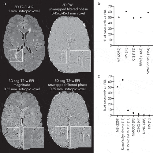
Figure 1. (A) Representative 3-tesla MRI example of a paramagnetic rim lesion (PRL) in a 21-year-old man with relapsing MS. PRL is clearly seen on both 3D seg-T2*-w EPI magnitude and unwrapped filtered phase images and on axial SWI phase images. For each image modality, PRL magnification is shown in the white insets. For each MRI sequence, image resolution is provided. (B) Graph showing the overall percentage of MS cases with at least one PRL by MS clinical phenotypes (number of cases evaluated in the brackets). (C) Graph showing the overall percentage of cases with at least one PRL by diseases (number of cases evaluated in the brackets).

Table 1. Clinical MRI studies assessing the diagnostic specificity and sensitivity of PRL in differentiating between MS and MS-mimic disorders.