Figures & data
Figure 1 SDS-polyacrylamide gel electrophoresis of G-6PD purified by affinity gel. Lane 1 and 2 are standard albumin (66.000) and lanes 3–4 are G-6PD.
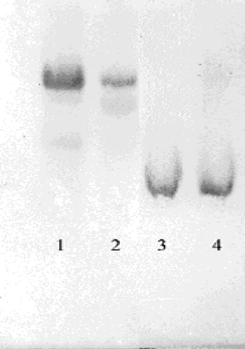
Figure 2 Activity % -[Omeprazole] regression analysis graphs for human erythrocytes G-6PD in the presence of 5 different omeprazole concentrations.
![Figure 2 Activity % -[Omeprazole] regression analysis graphs for human erythrocytes G-6PD in the presence of 5 different omeprazole concentrations.](/cms/asset/4268a14e-16bd-45a6-b746-18f68285b007/ienz_a_135662_f0002_b.gif)
Figure 3 Activity % -[Morphine Sulphate] regression analysis graphs for human erythrocytes G-6PD in the presence of 5 different morphine sulphate concentrations.
![Figure 3 Activity % -[Morphine Sulphate] regression analysis graphs for human erythrocytes G-6PD in the presence of 5 different morphine sulphate concentrations.](/cms/asset/569a6864-eca9-4ad5-ac70-d83917428dc4/ienz_a_135662_f0003_b.gif)
Figure 4 Activity % -[Vankomycin] regression analysis graphs for human erythrocytes G-6PD in the presence of 5 different vankomycin concentrations.
![Figure 4 Activity % -[Vankomycin] regression analysis graphs for human erythrocytes G-6PD in the presence of 5 different vankomycin concentrations.](/cms/asset/584dda9f-3062-4a3e-8f05-8696c08a5450/ienz_a_135662_f0004_b.gif)
Figure 5 Activity % -[Ketamine] regression analysis graphs for human erythrocytes G-6PD in the presence of 5 different ketamine concentrations.
![Figure 5 Activity % -[Ketamine] regression analysis graphs for human erythrocytes G-6PD in the presence of 5 different ketamine concentrations.](/cms/asset/5ea5ce13-46b4-42b2-b1f9-1296d82e7eb5/ienz_a_135662_f0005_b.gif)
Figure 6 Activity % -[Remifentanyl] regression analysis graphs for human erythrocytes G-6PD in the presence of 5 different remifentanyl concentrations.
![Figure 6 Activity % -[Remifentanyl] regression analysis graphs for human erythrocytes G-6PD in the presence of 5 different remifentanyl concentrations.](/cms/asset/d5859a1f-d2de-4d3d-850a-5938b916204b/ienz_a_135662_f0006_b.gif)
Figure 7 Lineweaver–Burk graphs for G-6PD in presence of three constant omeprazole and five different substrate concentrations.
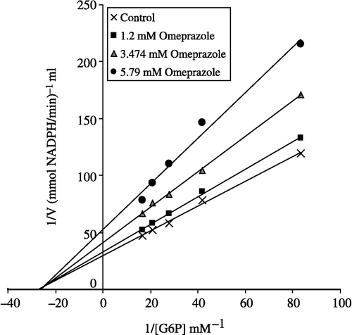
Figure 8 Lineweaver–Burk graphs for G-6PD in presence of three constant morphine sulphate concentrations and five different substrate concentrations.
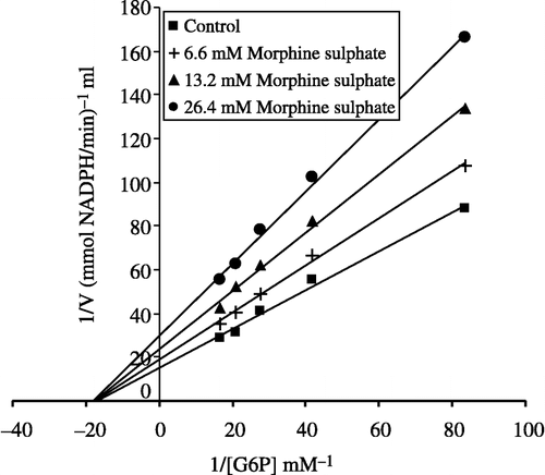
Figure 9 Lineweaver–Burk graphs for G-6PD in presence of three constant vankomycin concentrations and five different substrate concentrations.
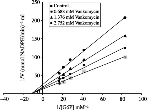
Table I. Purification scheme of glucose 6-phosphate dehydrogenase from human erythrocytes.
Table II. Ki and I50 values obtained from Lineweaver–Burk graphs for G-6PD in the presence of three fixed inhibitors and five substrate concentrations for omeprazole, morphine sulphate, and vankomycin. I50 values for remifentanyl and ketamine.