Figures & data
Scheme 1 Synthesis of compounds 1a–f.
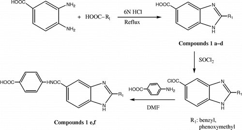
Table I. Physical properties of compounds 1a–f.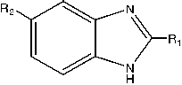
Figure 1 Dose-dependent inhibitory effect of benzimidazoles on H2O2-induced lipid peroxidation in erythrocyte membranes. Data are expressed as mean ± SE of 3–8 incubations.
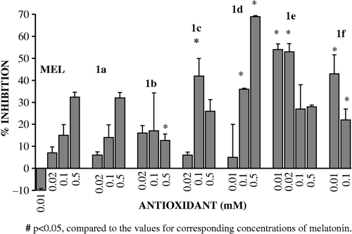
Figure 2 In vitro effect of melatonin and benzimidazole derivatives on SOD activity from human erythrocytes. 1a-f was added at 0.5mM concentration to the incubation media. Values represent means from at least 3 different incubations ± SEM.
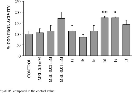
Figure 3 In vitro effect of melatonin and benzimidazole derivatives on catalase activity from human erythrocytes. 1a-d was added at 0.5mM, 1e at 0.02mM and 1f at 0.01mM concentration to the incubation media. Values represent means from at least 3 different incubations ± SEM.
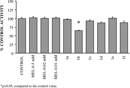
Figure 4 In vitro effect of melatonin and benzimidazole derivatives on G6PD activity from human erythrocytes. 1a-d was added at 0.5mM, 1e at 0.02mM and 1f at 0.01mM concentration to the incubation media. Values represent means from at least 3 different incubations ± SEM.
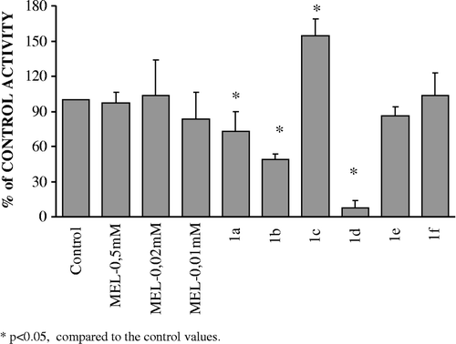
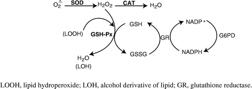
Figure 5 The interrelationships between antioxidant enzymes in detoxification of reactive oxygen species.