Figures & data
Table I. IC50 values against eukaryotic DNA topoisomerase I and II for the tested compounds and the reference drugs (μM) .
.
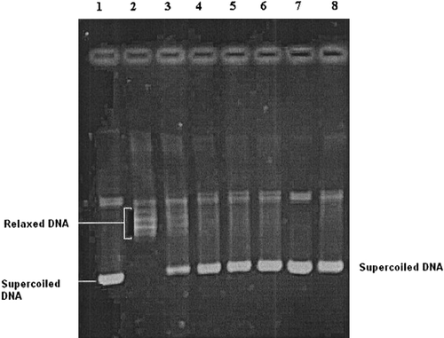
Figure 1 Electrophoregram showing the inhibitory effects of tested compound 17 and reference drug camptothecin on eukaryotic DNA topoisomerase I. Lane 1; supercoiled pBR322 plasmid DNA (0.3 μg) with incubation mixture without enzyme. Lane 2; plasmid DNA with 1 U of topo I enzyme (control). Lanes 3–6; plasmid DNA with 1 U of topo I enzyme in the presence of compound 17 at concentrations 0.1, 1, 5 and 10 μg/μL, respectively. Lane 7; plasmid DNA with compound 17 at a concentration of 10 μg/μL without enzyme. Lane 8; plasmid DNA with 1 U of topo I enzyme and a known DNA topoisomerase I inhibitor (camptothecin) at a concentration of 5 μg/μL. The relaxation assay in a cell-free system was performed as described in methods.
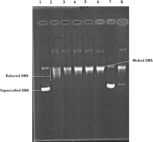
Figure 2 Electrophoregram showing the inhibitory effects of tested compound 6 and reference drug etoposide on eukaryotic DNA topoisomerase II. Lane 1; supercoiled pBR322 plasmid DNA (0.3 μg) with incubation mixture without enzyme. Lane 2; plasmid DNA with 5 U of topo II enzyme (control). Lanes 3–6; plasmid DNA with 5 U of topo II enzyme in the presence of compound 6 at concentrations 0.1, 1, 5 and 10 μg/μL, respectively. Lane 7; plasmid DNA with compound 6 at a concentration of 10 μg/μL without enzyme. Lane 8; plasmid DNA with 5 U of topo II enzyme and a known DNA topoisomerase II inhibitor (etoposide) at a concentration of 10 μg/μL. The relaxation assay in a cell-free system was performed as described in methods.