Figures & data
Figure 1. The chemical structure of cyanidin-3-galactoside.
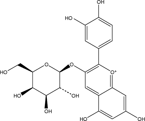
Figure 2. The percentage enzyme inhibition of acarbose and its combination with cyanidin-3-galactoside(Cya3Gal) on intestinal α-glucosidase (sucrase). Results were expressed as mean ± S.E.M., n = 3. * P < 0.01 compared to acarbose alone.
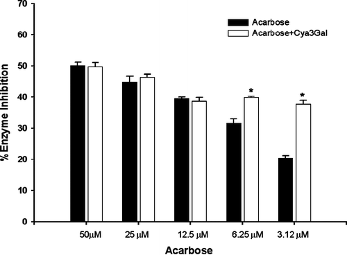
Figure 3. The percentage enzyme inhibition of acarbose and its combination with cyanidin-3-galactoside(Cya3Gal) on intestinal α-glucosidase (maltase). Results were expressed as mean ± S.E.M., n = 3. * P < 0.01 compared to acarbose alone.
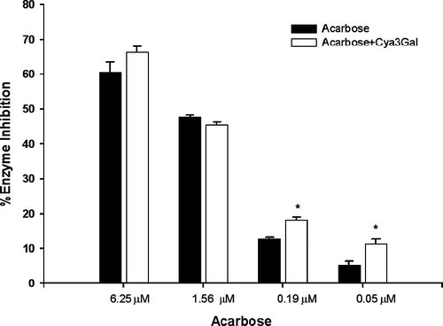
Figure 4. (A) The data were presented in a Lineweaver–Burk plot, 1/v against l/[S]. (B) Secondary replot of slope vs. [I] from a primary Lineweaver–Burk plot for the determination of Ki. The value of R2 and slope were 0.990 (C) Secondary replot of intercept vs. [I] from a primary Lineweaver–Burk plot for the determination of Ki′. The value of R2 and slope were 0.991.
![Figure 4. (A) The data were presented in a Lineweaver–Burk plot, 1/v against l/[S]. (B) Secondary replot of slope vs. [I] from a primary Lineweaver–Burk plot for the determination of Ki. The value of R2 and slope were 0.990 (C) Secondary replot of intercept vs. [I] from a primary Lineweaver–Burk plot for the determination of Ki′. The value of R2 and slope were 0.991.](/cms/asset/46fe159b-71c0-44e9-93d4-eb893b59b037/ienz_a_290860_f0004_b.gif)