1,281
Views
23
CrossRef citations to date
0
Altmetric
Research Article

Green tea catechins in chemoprevention of cancer: A molecular docking investigation into their interaction with glutathione S-transferase (GST P1-1)

, , , &
Pages 287-295 | Received 18 Dec 2007, Accepted 28 Feb 2008, Published online: 01 Feb 2009

Figures & data

Figure 1 . Representative scheme of the glutathione pathway and ABC-transporters family role in drug elimination and defence. Glutathione S-transferases (GSTs) conjugate GSH to drugs and drug metabolites facilitating the ATP-dependent elimination of drugs or drug-GS conjugates, involving ABC transporters (like ABCB1, ABCC1 and ABCG2). The drug/drug metabolites and intracellular GSH equilibrium is a major determinant of blood drug level.
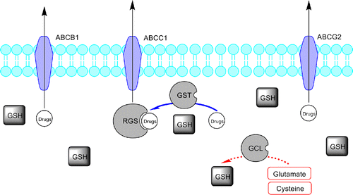
Figure 1 . Representative scheme of the glutathione pathway and ABC-transporters family role in drug elimination and defence. Glutathione S-transferases (GSTs) conjugate GSH to drugs and drug metabolites facilitating the ATP-dependent elimination of drugs or drug-GS conjugates, involving ABC transporters (like ABCB1, ABCC1 and ABCG2). The drug/drug metabolites and intracellular GSH equilibrium is a major determinant of blood drug level.

Figure 2 . Chemical structures of the green tea catechins and of the alkylating anti-cancer drugs docked. The oxygen numbering scheme and the ring nomenclature [Citation43] used throughout the text is indicated only for ( − )-EGCG.

Figure 2 . Chemical structures of the green tea catechins and of the alkylating anti-cancer drugs docked. The oxygen numbering scheme and the ring nomenclature [Citation43] used throughout the text is indicated only for ( − )-EGCG.

Figure 3 . Left: binding into hGST P1-1 of ( − )-EGCG red, ( − )-GCG yellow, ( − )-EGC blue and ( − )-CG green. Right: binding into hGST P1-1of cyclophosphamide gray, ifosfamide light blue, melphalan pink and chlorambucil gold. The protein is represented as a light grey Connolly surface, while the ligands as stick models.

Figure 3 . Left: binding into hGST P1-1 of ( − )-EGCG red, ( − )-GCG yellow, ( − )-EGC blue and ( − )-CG green. Right: binding into hGST P1-1of cyclophosphamide gray, ifosfamide light blue, melphalan pink and chlorambucil gold. The protein is represented as a light grey Connolly surface, while the ligands as stick models.

Table I.  Heavy atoms (HA), molecular weight (MW) and QM/MM interaction energies (kcal/mol) for the eight studied compounds.

Figure 4 . Left: 3D structure of hGST P1-1 (solid ribbon) in complex with ( − )-EGCG (top) and ( − )-GCG (bottom). Residues lining the ligand position are represented as a white Connolly surface, while the ligands are represented by a green Connolly surface. Right: binding mode of ( − )-EGCG (top) and ( − )-GCG (bottom) within hGST P1-1. Ligand (CPK) and the residues involved in interactions (orange, labelled) are represented in stick while the hydrogen bonds are shown as green dotted lines.
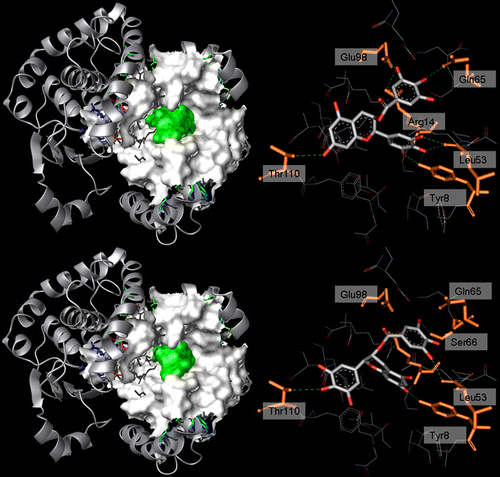
Figure 4 . Left: 3D structure of hGST P1-1 (solid ribbon) in complex with ( − )-EGCG (top) and ( − )-GCG (bottom). Residues lining the ligand position are represented as a white Connolly surface, while the ligands are represented by a green Connolly surface. Right: binding mode of ( − )-EGCG (top) and ( − )-GCG (bottom) within hGST P1-1. Ligand (CPK) and the residues involved in interactions (orange, labelled) are represented in stick while the hydrogen bonds are shown as green dotted lines.

Figure 5 . Binding within hGST P1-1 of the considered drugs: cyclophosphamide (a), ifosfamide (b), chlorambucil (c) and melphalan (d). Ligand (CPK) and interacting key residues (orange) are represented as stick models, hydrogen bonds are shown as green dotted lines.
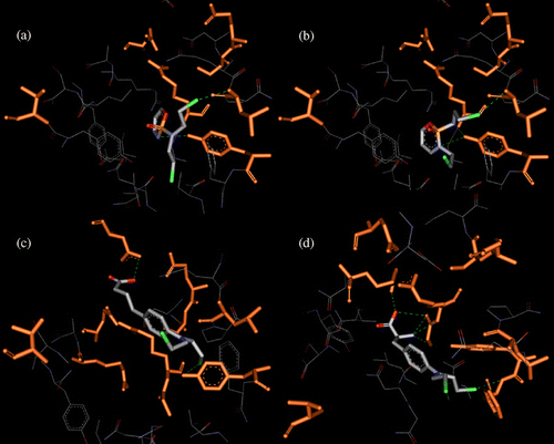
Figure 5 . Binding within hGST P1-1 of the considered drugs: cyclophosphamide (a), ifosfamide (b), chlorambucil (c) and melphalan (d). Ligand (CPK) and interacting key residues (orange) are represented as stick models, hydrogen bonds are shown as green dotted lines.

Reprints and Corporate Permissions

Please note: Selecting permissions does not provide access to the full text of the article, please see our help page How do I view content?

To request a reprint or corporate permissions for this article, please click on the relevant link below:

Academic Permissions

Please note: Selecting permissions does not provide access to the full text of the article, please see our help page How do I view content?

Obtain permissions instantly via Rightslink by clicking on the button below:

If you are unable to obtain permissions via Rightslink, please complete and submit this Permissions form. For more information, please visit our Permissions help page.