Figures & data
Figure 1. A: IAP activity before and after EGTA addition. B: IAP activity with different calcium concentrations obtained after calcium addition to IAP that was previously treated with EGTA 50 mmole/L. C: Western blot of IAP before (lane 1) and after EGTA addition (lane 2) and after 50 mmole/L calcium addition (lane 3). Data are expressed as mean ± SD (n = 4).
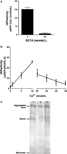
Table I. Molecular mass and isoelectric point obtained by 2D-electrophoresis for the dimer and the aggregated form with and without calcium addition.
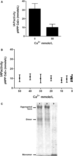
Figure 2. A: IAP activity before and after calcium addition. B: IAP activity as a function of calcium concentration. Calcium concentrations of the horizontal axis are obtained after different additions of EGTA to the solution that started with 50 mmole Ca/L. C: Western blot of IAP before (lane 1) and after Ca2 + addition (lane 2) and after EGTA addition (lane 3) to obtain 0 mmole/L free calcium. Data are expressed as mean ± SD (n = 4).