Figures & data
Figure 1. The polyol pathway of glucose metabolism.
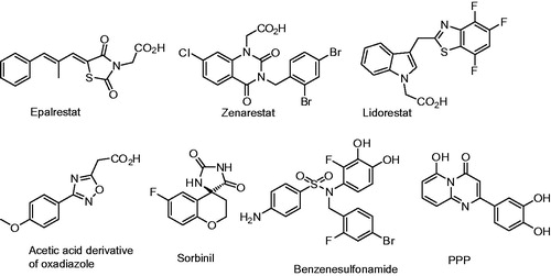
Figure 2. Previously reported aldose reductase inhibitors.
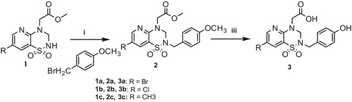
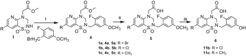
Scheme 1. Reagents and conditions: (i) K2CO3, (ii) CH3CN, 70 °C; (iii) BBr3, CH2Cl2, 0 °C.
Scheme 2. Reagents and conditions: (i) K2CO3, CH3CN, 70 °C; (ii) NaOHaq., THF, then HClaq. and (iii) K2CO3, thiophenol, NMP, 150 °C.
Scheme 3. Reagents and conditions: (i) K2CO3, CH3CN, 70 °C; (ii) AlCl3, CH2Cl2, 40 °C and (iii) NaOHaq., THF, then HCaq.
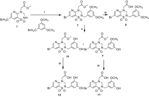
Table 1. Biological evaluation of pyridothiadiazine derivatives.
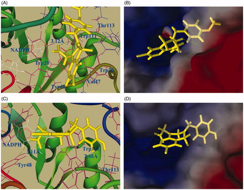
Figure 3. Molecular docking of compound 13b (A & B) and 3b (C & D). (A & C) Docking of compounds 13b or 3b into the ALR2 active site. The protein structure is shown in ribbon and tube representation with selected residues labeled and shown in line representation; ligand 13b (or 3b) and NADPH are shown as stick models. The docked pose of 13b and 3b are shown in yellow. Hydrogen bonds are shown as green dashed lines. (B & D) Surface representation of protein active site residues in the docking of 13b or 3b into the active site of ALR2.