Figures & data
Figure 1. Examples of colchicine binding site inhibitors.
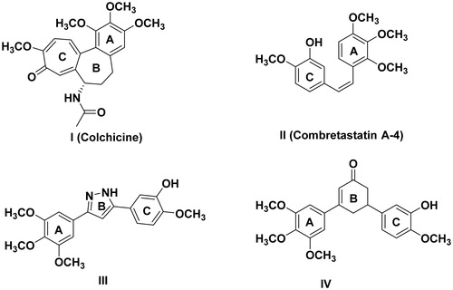
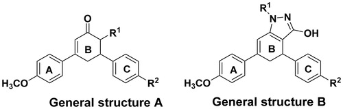
Figure 2. General structures of target compounds.
Scheme 1. Synthesis of compounds 3a,b, 4a,b and 5a–f.
Scheme 2. Synthesis of compounds 6a,b, 7a,b, 8a,b and 9a–f.
Figure 3. Tautomeric forms of 1H-indazol-3-ol.
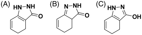
Table 1. Antiproliferative activity against HCT-116 cell line and MCF-7.
Table 2. Percentage inhibition of tubulin polymerization.
Figure 4. Cell cycle analysis histograms for HCT-116 cells. (A) Control, (B) 4b, (C) 5e and (D) 5f.
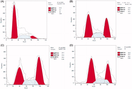
Figure 5. Cell cycle analysis histograms for MCF-7 cells. (A) Control, (B) 4b, (C) 5e and (D) 5f.
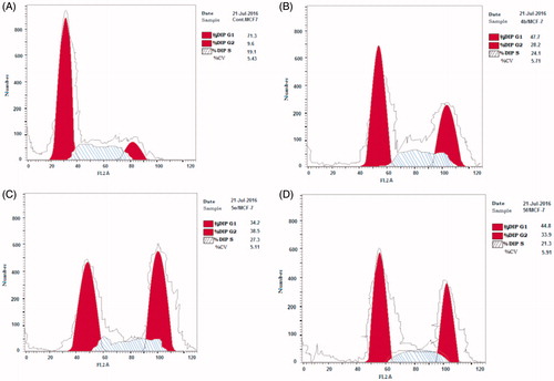
Table 3. Results of cell cycle analysis in HCT-116 and MCF-7 for compounds 4b, 5e and 5f.
Table 4. Results of the molecular docking study.
Figure 6. 2D interaction diagram of the top docking pose of the R isomer of compound 4b.
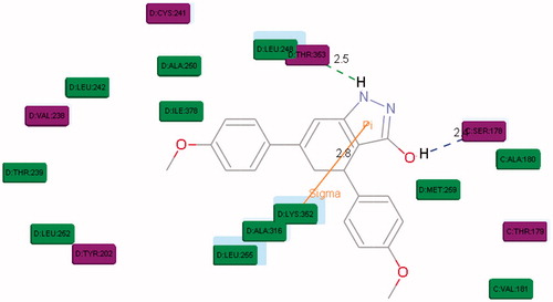
Figure 7. Overlay of the top docking poses of R (green) and S (yellow) isomers of 4b in the active site of tubulin (PDB: 1SA0).
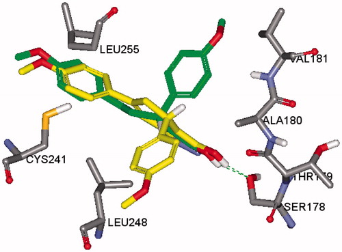
Figure 8. 2D interaction diagram of the top docking pose of the R isomer of compound 5e.
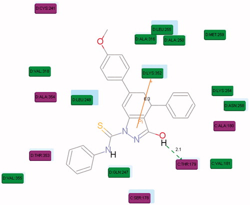
Figure 9. 2D interaction diagram of the top docking pose of the R isomer of compound 5f.
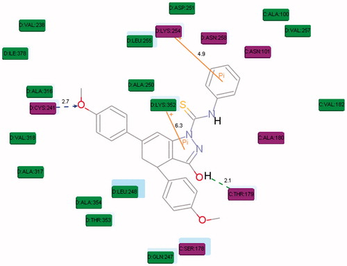
Figure 10. 2D interaction diagram of the top docking pose of the S isomer of compound 5e.
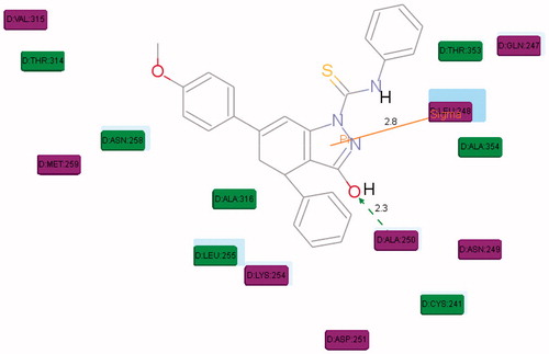
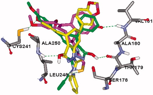
Figure 11. Overlay of the top docking poses of R (green), S (yellow) isomers of 5e and DAMA-colchicine (magenta) in the active site of tubulin (PDB: 1SA0).