Figures & data
Figure 1. (a) Schematic structure of non-peptide inhibitors bearing the 2-cloronaphthoquinonic unit. (b) The general structure of dipeptide derivatives with a 2-chloronaphthoquinone group. (c) The generic structure of the new amino acid derivatives linked to the 2-chloronaphthoquinone group.
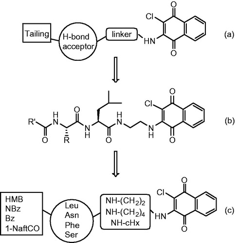
Table 1. Inhibition of the proteasome subunits by the synthesised compounds.
Scheme 1. Synthesis of naphthoquinone amino acid derivatives.
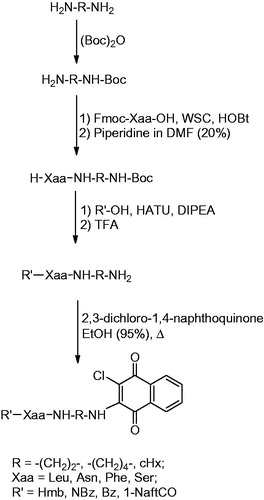
Figure 2. Effect of compounds 4, 6, 9–11 on cell proliferation. (a) MDA and (b) A2780 tumour cells cultured for 3 d in the presence or absence of the indicated concentrations of compounds. The means of three independent experiments performed in duplicate are shown.
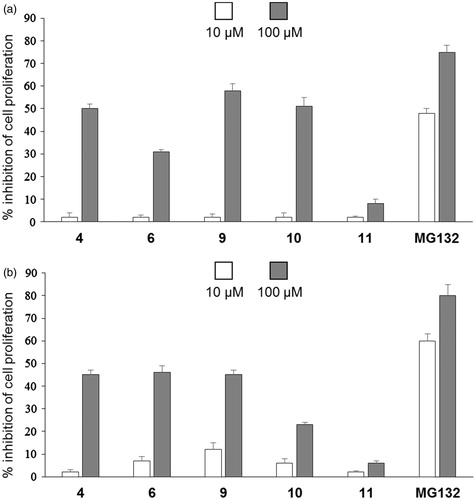
Figure 3. (a) Molecule 10 in the β1 active site (best pose). (b) Schematic view of the interactions between the receptor and the docked molecule.
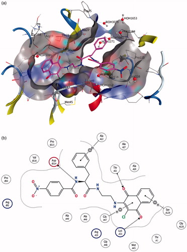
Figure 4. (a) Molecule 10 in the β5 active site. (b) Schematic view of the interactions between the receptor and the docked molecule.
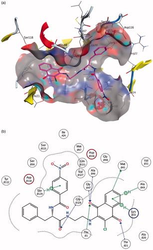
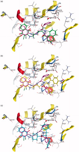
Figure 5. Molecule 16 (a), 26 (b) and 42 (c) docked into the ?1 binding site. Molecule 10 docked in the same binding site is reported for comparison.
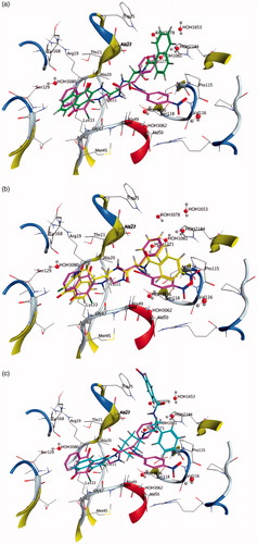
Figure 6. Molecule 16 (a), 26 (b) and 42 (c) docked into the β5 binding site. Molecule 10 docked in the same binding site is reported for comparison.