
Figures & data
Figure 1. Structures of VEGFR inhibitors.
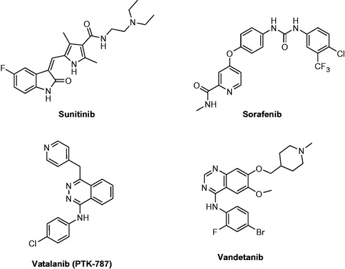
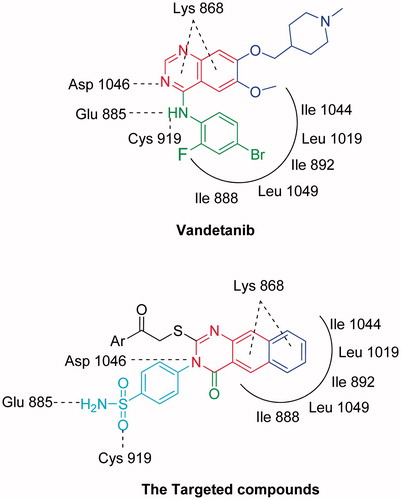
Figure 2. The design concepts of the targeted compounds.
Scheme 1. Formation of benzoquinazoline-sulfonamide derivatives 3–12.
Scheme 2. Formation of benzoquinazoline-sulfonamide derivatives 13–19.
Scheme 3. Formation of benzoquinazoline-sulfonamide derivatives 18–24.
Table 1. VEGFR-2 inhibitory activity and anti-proliferative activity against MCF-7 cell line.
Figure 3. The effect of compound 9 and vandetanib on the activation of caspase-3 in MCF-7 cells.
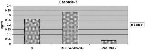
Table 2. Effect of compound 9 on the active caspases-3 in MCF-7 cells.
Figure 4. The effect of compound 9 and vandetanib on the level of Bax and BCl2.
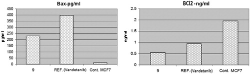
Table 3. Effect of compound 9 on the expression of the gene of some apoptosis key markers.
Figure 5. The effect of compound 9 and vandetanib on the cell cycle phases.
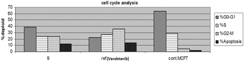
Figure 6. The effect on the phases of the cell cycle (A) compound 9, (B) Vandetanib, (C) control MCF-7 cells.
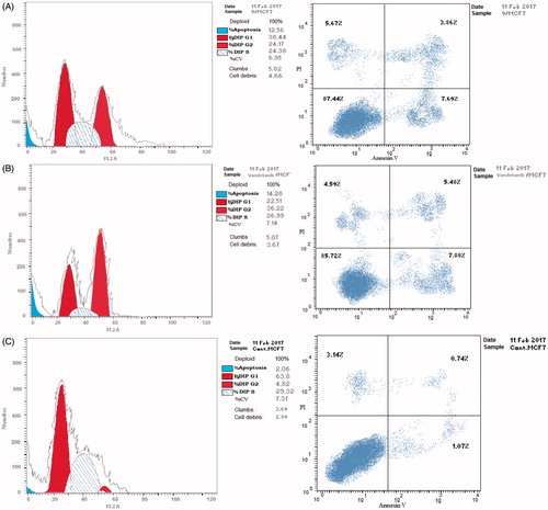
Figure 7. Cytotoxic activity of compound 9 and vandetanib towards MCF-12 A normal breast cell line.
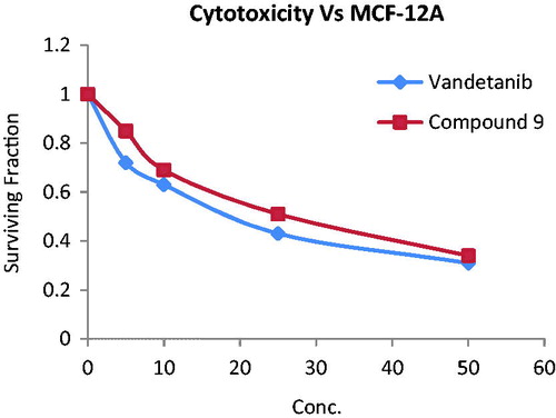
Figure 8. 2D interaction of the co-crystallised ligand inside the active site of 3U6J.
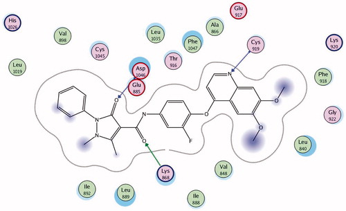
Figure 9. 2D interaction of compound 9 inside the active site of 3U6J.
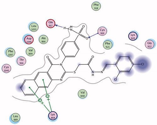
Figure 10. 3D interaction of compound 9 inside the binding site of 3U6J.
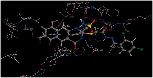
Figure 11. 2D interaction of compound 22 inside the active site of 3U6J.
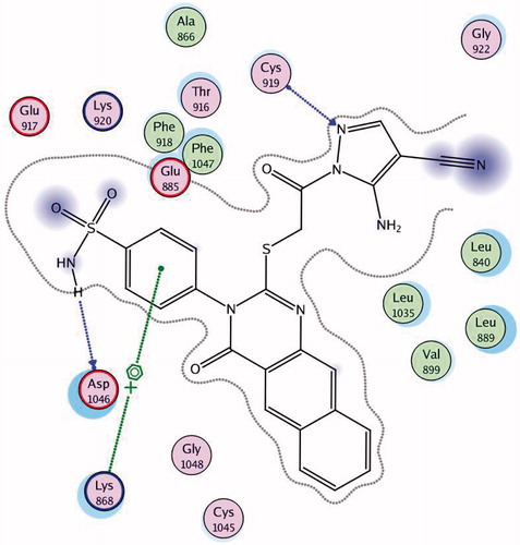
Figure 12. 3D interaction of compound 22 inside the binding site of 3U6J.
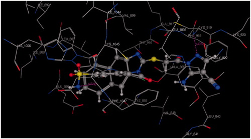
Table 4. Docking results of the promising compounds inside the 3U6J active site.