Figures & data
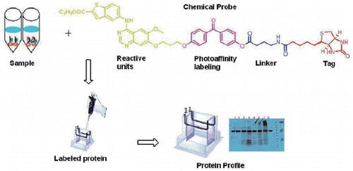
Figure 1. Structures and protein labelling of photoaffinity probes.
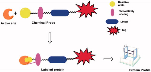
Figure 2. Design of the novel photoaffinity probe.
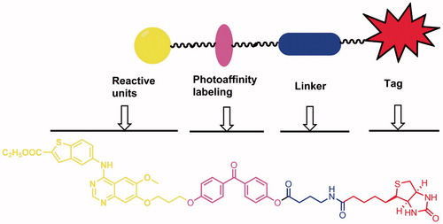
Scheme 1. The preparation of the photoaffinity probe. Reagents and conditions: (a) 1-bromo-3-chloropropane, Potassium carbonate, DMF, 70 °C; (b) Nitric acid, acetic acid, acetic anhydride, 0–5 °C; (c) Pd/C, methanol, rt; (d) Formamidine acetate, alcohol, reflux; (e) thionyl chloride, DMF, reflux; (f) ethyl 5-aminobenzo[b]thiophene-2-carboxylate, isopropanol, reflux; (g) 4,4-dihydroxybenzophenone (DHBP), Potassium carbonate, DMF, 60 °C; (h) γ-aminobutyric acid, isobutyl chlorocarbonate, DMF, rt; (i) EDCI/DMAP, DMF, rt.
![Scheme 1. The preparation of the photoaffinity probe. Reagents and conditions: (a) 1-bromo-3-chloropropane, Potassium carbonate, DMF, 70 °C; (b) Nitric acid, acetic acid, acetic anhydride, 0–5 °C; (c) Pd/C, methanol, rt; (d) Formamidine acetate, alcohol, reflux; (e) thionyl chloride, DMF, reflux; (f) ethyl 5-aminobenzo[b]thiophene-2-carboxylate, isopropanol, reflux; (g) 4,4-dihydroxybenzophenone (DHBP), Potassium carbonate, DMF, 60 °C; (h) γ-aminobutyric acid, isobutyl chlorocarbonate, DMF, rt; (i) EDCI/DMAP, DMF, rt.](/cms/asset/409b741a-ef5c-4f67-8567-9abd29526ed7/ienz_a_1344979_sch0001.gif)
Figure 3. Photoaffinity labelling of cell lysate by synthesised photoaffinity probe. Lane 1: whole cell lysate; Lane 2: DMSO treatment; Lane 3: 5 μM probe treatment; Lane 4: 10 μM probe treatment; Lane 5: 5 μM probe treatment plus UV exposure; Lane 6: 10 μM probe treatment plus UV exposure. MIAPaCa2 cell lysate was treated as indicated and pull-down assay was then performed using streptavidin agarose. The samples were then run western blot and probed with streptavidin-HRP antibody. Arrows point out specific bands of streptavidin blot. * Non-specific bands.
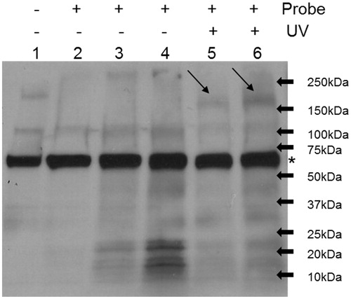
Figure 4. Synthesised photoaffinity probe binds to EGFR and HER2 proteins. Lane 1: whole cell lysate; Lane 2: DMSO treatment; Lane 3: 10 μM probe and 100 μM compound 17 co-treatment; Lane 4: 10 μM probe treatment; Lane 5: DMSO treatment plus UV exposure; Lane 6: 10 μM probe treatment plus UV exposure; Lane 7: 10 μM probe and 100 μM compound 17 co-treatment under UV exposure. MIAPaCa2 cell lysate was treated as indicated and pull-down assay was then performed using streptavidin agarose. The samples were then run western blot and probed with anti-EGFR (left) or anti-HER2 (right) antibody.
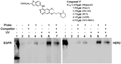
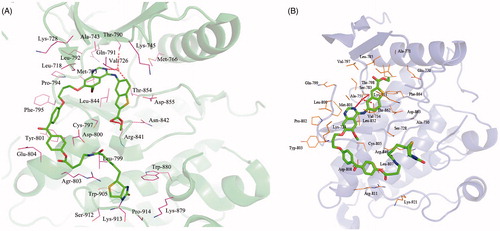
Figure 5. Binding models of photoaffinity probe target into active site of EGFR (A) and HER2 (B).