Figures & data
Scheme 1. 2,3-Substituted-quinoxaline and 1,4-substituted-[1,2,4]triazolo[4,3-a]quinoxalin derivatives.
![Scheme 1. 2,3-Substituted-quinoxaline and 1,4-substituted-[1,2,4]triazolo[4,3-a]quinoxalin derivatives.](/cms/asset/0ba9a270-ce73-420e-8660-b83e335bbd7c/ienz_a_1363743_sch0001.jpg)
Figure 1. Chemical structures of series 3, series 4, and quercetin.
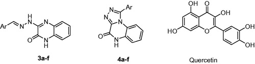
Table 1. Inhibitory activity of the title compounds against isoforms of sPLA2 expressed as IC50 (µM)±standard deviation.
Table 2. Inhibitory activity of the title compounds against α-glucosidase and α-amylase expressed as IC50 (µM) ± standard deviation.
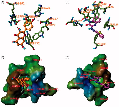
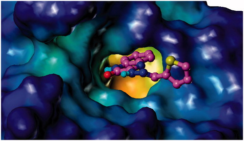
Figure 2. Binding mode of 3f and 4c to hG-X sPLA2. (A) Compound 3f (magenta) and (C) compound 4c (orange) in the binding pocket of hG-X sPLA2 showing hydrogen bonds, calcium coordination (purple), and interacting residues. (B) Compound 3f (magenta) and (D) compound 4c (orange) in the binding pocket of hG-X sPLA2 showing a lipophilic potential surface of the pocket.
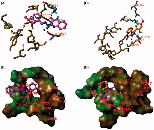
Figure 3. Binding mode of quercetin and 3e to α-glucosidase. (A) Quercetin (orange) and (C) compound 3e (magenta) in the binding pocket of α-glucosidase showing hydrogen bonds and interacting residues. (B) Quercetin (orange) and (D) compound 3e (magenta) in the binding pocket of α-glucosidase showing a lipophilic potential surface of the pocket.