1,854
Views
101
CrossRef citations to date
0
Altmetric
Research Paper

Carbon- versus sulphur-based zinc binding groups for carbonic anhydrase inhibitors?

Pages 485-495 | Received 07 Jan 2018, Accepted 12 Jan 2018, Published online: 02 Feb 2018

Figures & data

Figure 1. Clinically used sulphonamides/sulphamates with CA inhibitory action (A–S) and SLC-0111 (T) in phase II clinical trials as an antitumour/antimetastatic agentCitation18,Citation19.

Figure 1. Clinically used sulphonamides/sulphamates with CA inhibitory action (A–S) and SLC-0111 (T) in phase II clinical trials as an antitumour/antimetastatic agentCitation18,Citation19.

Figure 2. General scheme showing CAIs belonging to the zinc binders class (sulphonamides, sulphamates, sulphamides, carboxylates, hydroxamates, phosphonates, borols, etc.) in interaction with an α-CACitation1,Citation3. The ZBG is coordinated to the metal ion and makes hydrogen bonds with the gate keeper residues Thr199–Glu106, conserved in all α-CAsCitation1–3. The scaffold of the inhibitor may occupy either the hydrophylic or hydrophobic (or both) halves of the active siteCitation12, whereas the tails are orientated towards the exit of the cavity where the most variable amino acid residues among the different mammalian CAs are locatedCitation26–28. The interactions between the scaffold/tail with the enzyme are not shown.

Figure 2. General scheme showing CAIs belonging to the zinc binders class (sulphonamides, sulphamates, sulphamides, carboxylates, hydroxamates, phosphonates, borols, etc.) in interaction with an α-CACitation1,Citation3. The ZBG is coordinated to the metal ion and makes hydrogen bonds with the gate keeper residues Thr199–Glu106, conserved in all α-CAsCitation1–3. The scaffold of the inhibitor may occupy either the hydrophylic or hydrophobic (or both) halves of the active siteCitation12, whereas the tails are orientated towards the exit of the cavity where the most variable amino acid residues among the different mammalian CAs are locatedCitation26–28. The interactions between the scaffold/tail with the enzyme are not shown.

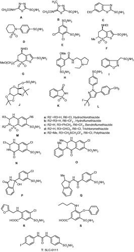
Table 1. Inhibition of CA isozymes I–XV (of human = h, and murine = m origin) with compounds 110.

Figure 3. Superimposition of the hCA II – 2 adduct (blue, PDB code 2WEJCitation42) with the hCA II – 4 adduct (violet, PDB code 3T5UCitation37), the hCA II – 5 adduct (silver, PDB code 3T5ZCitation37) and the hCA II – hydroxamate 9 adduct (green, PDB code 4FL7Citation38).

Figure 3. Superimposition of the hCA II – 2 adduct (blue, PDB code 2WEJCitation42) with the hCA II – 4 adduct (violet, PDB code 3T5UCitation37), the hCA II – 5 adduct (silver, PDB code 3T5ZCitation37) and the hCA II – hydroxamate 9 adduct (green, PDB code 4FL7Citation38).

Table 2. hCA I, II, IX, and XII inhibition data with compounds 2, 2a, 2b, 7, and 7a.