Figures & data
Figure 1. Structures of bile acids 1–10.
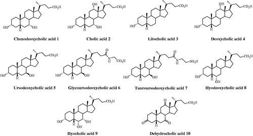
Figure 2. Structures of steroids 11–22.
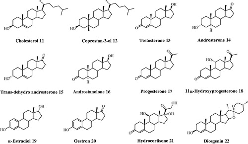
Table 1. Inhibition data of human CA isoforms hCA I, II, IV and IX with compounds reported here and the standard sulfonamide inhibitor acetazolamide (AAZ) by a stopped flow CO2 hydrase assay.
Figure 3. Dockings of (A) α-estradiol (19) and (B) tauroursodeoxycholic acid (7) within hCA II. (C) Superposed docked hyocholic acid (9) (blue) and cholic acid (2) (yellow) X-ray solved orientation within hCA II.
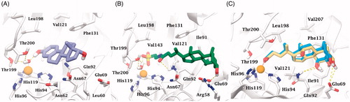
Figure 4. Analysis of the MD simulation of 9 docked to hCA II. (A) Coordination and H-bonds occupancies within 10 ns MD for 9 - hCA II complex. (B) Rmsd representation of the heavy atoms of the receptor and the ligand from the starting model structure during the simulation.
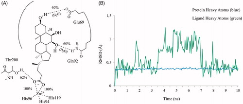
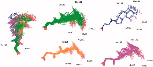
Figure 5. Superposed representative orientations of the four identified clusters within superposed protein backbones of 200 frames of MD.
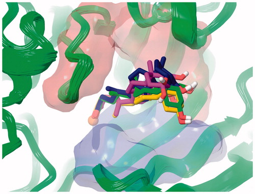
Figure 6. Conformer families of 9 identified over the 10 ns MD period.