Figures & data
Figure 1. Three of the most studied DGKα inhibitors.
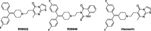
Figure 2. Structures of the deaza analogue of R59949 and Amb639752.
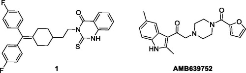
Figure 3. First set of compounds tested for their inhibitory activity on DGKα.
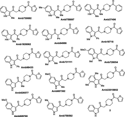
Table 1. Inhibitory activity on DGKα (I).
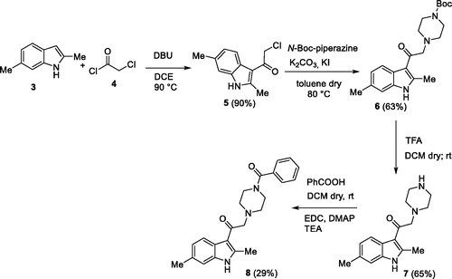
Scheme 1. The first synthetic route for the compound 8.
Scheme 2. The second synthetic route for the compound 8.
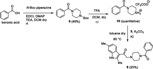
Figure 4. Carboxylic acids used.
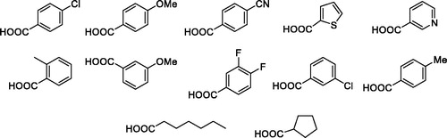
Figure 5. Putative DGKα inhibitors synthesised. In brackets the yield of the coupling reaction with the common intermediate 7.
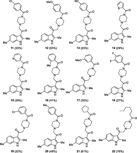
Table 2. Inhibitory activity on DGKα (II).
Figure 6. Dose–response curves for novel DGKα inhibitors. Dose–response of the most active compounds along with their IC50 values. Data from at least three independent experiments performed in triplicate.
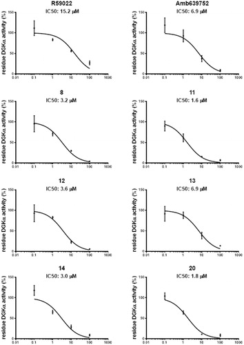
Figure 7. Isoform specificity of novel DGKα inhibitors. 293T cells were transfected with different DGK isoforms (A – DGKα, B – DGKζ, C – DGKθ, respectively) or empty vectors and homogenised. All the molecules were tested at 100 μM for their capacity to inhibit the DGK activity of the different isoform homogenates. Data are means ± SEM of at least three independent experiments performed in triplicate.
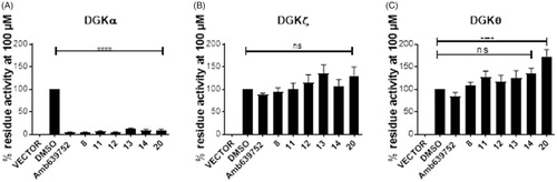
Figure 8. Novel DGKα inhibitors do not affect serotonin signalling. Human monocytes were pre-incubated for 1 h with the indicated drugs in absence or presence of serotonin and then stimulated with PMA 1 µM for 30 min (■ control unstimulated cells, ![]()
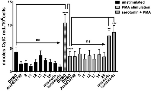
Figure 9. DGKα inhibitors partially restores apoptosis in SAP deficient lymphocytes. Lymphocytes from normal subjects were transfected with control or SAP specific siRNA (■ control siRNA, ■ SAP siRNA). After 4 days, the cells were restimulated with CD3 agonist OKT3 (10 ng/mL) in presence of respective inhibitor. Vehicle (DMSO) was used as control. Twenty-four hours later, the % of cell loss was evaluated by PI staining. Data are the mean ± SEM of nine independent experiments performed in triplicate.
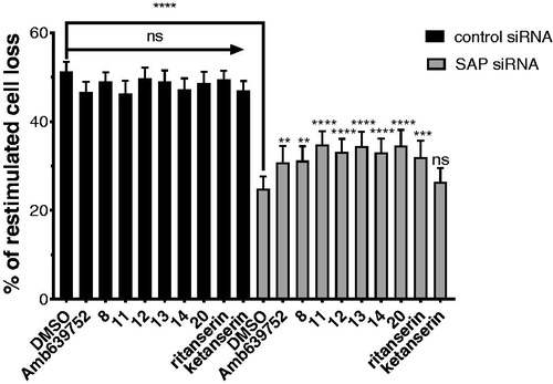
Figure 10. Novel DGKα inhibitors slow tumour cell migration. MCF7 monolayer was wounded and treated for 15 h with serum in presence of our new DGKα inhibitors (10 μM) or vehicle (DMSO). Results are expressed as the percentage of wound area compared to the initial area. Data are the mean ± SEM of nine independent experiments.
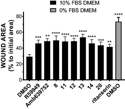
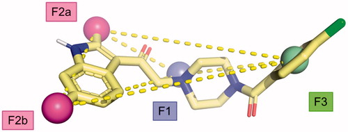
Figure 11. Proposed pharmacophoric model.