Figures & data
Figure 1. Chemical structures of well-known MAO-B inhibitors.
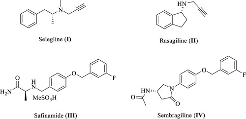
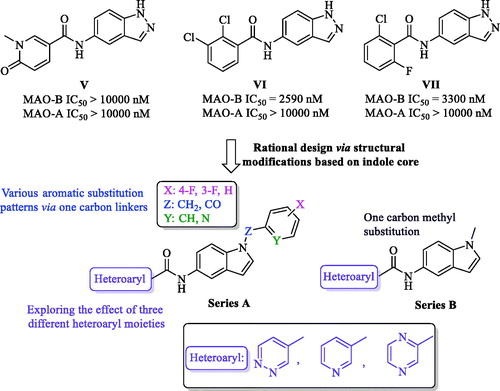
Figure 2. Rational design of the newly synthesized analogues (series A and B).
Scheme 1. Reagents and conditions: (a) appropriate halide derivative, NaH (60% in oil), DMF, 0–100 °C, 24 h; (b) dimethyl carbonate, K2CO3, DMF, reflux, 3 h; (c) NH4Cl, Fe, EtOH/H2O, reflux, 1 h; (d) appropriate heteroaryl carboxylic acid, DIPEA, HATU, DMF, MW, 116 °C, 45 min.
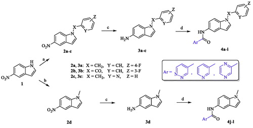
Table 1. Inhibitory effects of the synthesized compounds against MAO-B.
Figure 3. Dose dependent assay of compounds 4b and 4e over MAO-B.

Table 2. Inhibitory effects of the compounds 4b and 4e against MAO-A.
Figure 4. Type of inhibition of MAO-B by compound 4e. The catalytic rates were measured at different concentrations of benzylamine (0.065, 0.125, 0.25, 0.5, 1, 2, and 4 mM) in the absence and in the presence of different concentrations (10, 30, and 100 nM) of compound 4e. The Vmax, Km, and Ki values were calculated using SigmaPlot®.
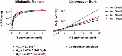
Figure 5. The docked model of the most active compounds 4b (A) and 4e (B) into MAO-B binding pocket.
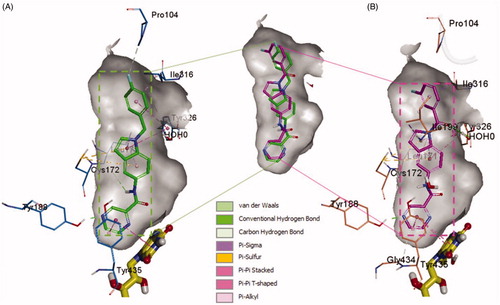
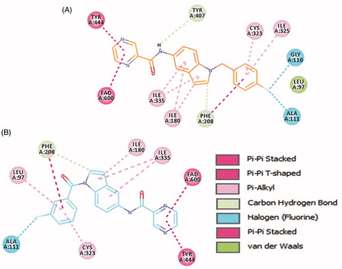
Figure 6. Two-dimensional (2D) interaction model for 4b (A) and 4e (B) with MAO-B.
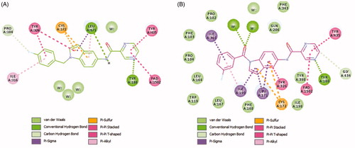
Figure 7. Binding orientation of ligand 4b and 4e inside the MAO-A protein structure. Ligand 4b (A) and 4e (B) shown in orange and blue colour stick format. FAD molecule shown in yellow stick format. Hydrophobic surface area shown around the ligand.
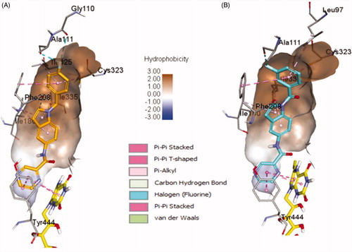
Figure 8. 2D interactions of ligands 4b and 4e inside MAO-A binding pocket. 4b (A) and 4e (B) ligands shown in orange and blue colour, respectively.