Figures & data

Figure 2. Representative dot-plots presenting the loss of mitochondrial membrane potential (MMP) in MCF-7 (A), and MDA-MB-231 (B) cells incubated with Epo (Epo100, 100 IU/ml) and LFM-A13 (LFM100, 100 μM) for 48 h (mean ± SD; n = 3). Cells with normal MMT are shown on the left side of the plots, cells with decreased MMT on the right side of the plots.
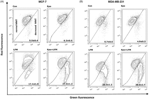
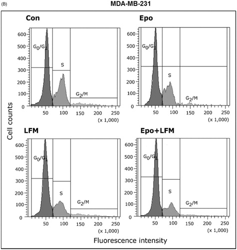
Figure 5. Representative examples of immunohistochemistry for BTK expression in breast cancer cells.
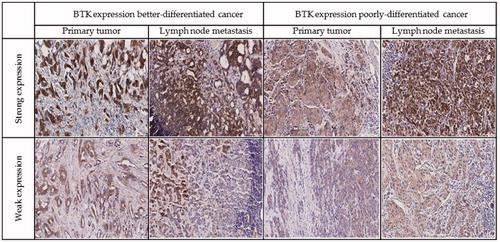
Table 1. BTK expression in primary tumours and metastases to lymph nodes.
Table 2. Spearman’s rho correlation between BTK level in tumour and node metastasis and selected parameters.
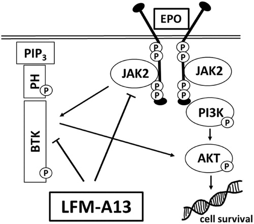
Figure 6. Schematic diagram of intracellular proteins BTK and JAK2 interaction and LFM-A13 mechanism of action. EPO: erythropoietin, LFM-A13: Bruton’s tyrosine kinase inhibitor; JAK2: non-receptor tyrosine kinase; BTK: Bruton’s tyrosine kinase; Akt: protein kinase B; PIP3: phosphatidylinositol-3,4,5-triphosphate; PH: pleckstrin homology domain.