Figures & data
Table 1. Types and subfamilies of topoisomerases
Figure 1. General mechanism of action of topoisomerase I (a) Top I binds to the DNA, (b) single-strand DNA (in blue) splitting, (c) controlled rotation of free DNA strand (in red), (d) religation of cleaved DNA strand.
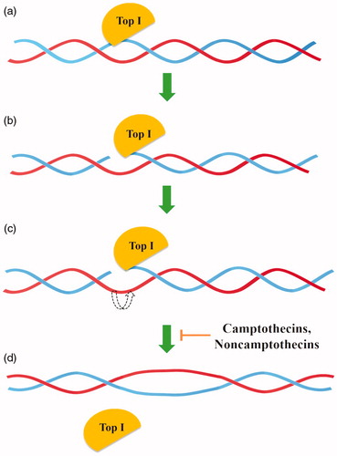
Figure 2. General mechanism of action of topoisomerase II (a) topoisomerase binds to the G-segment, (b) Top IIA- G-segment complex binds to T-segment, (c) Two ATP molecules are attached to the resulting complex, (d) G-segment cleavage in presence of Mg2+ ions, (e) T-segment transport through the created gap, (f) T-segment release and religation of G-segment broken strands and (g) hydrolysis of ATP molecules and release of the G-segment.
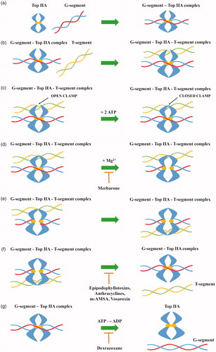
Table 2. Clinically relevant topoisomerases inhibitors
Table 3. Selected topoisomerase inhibitors under clinical investigation
Figure 3. Chemical structure of camptothecin.
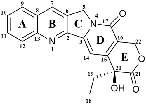
Figure 4. Chemical structures of camptothecins.

Figure 5. Chemical structures of noncamptothecins.
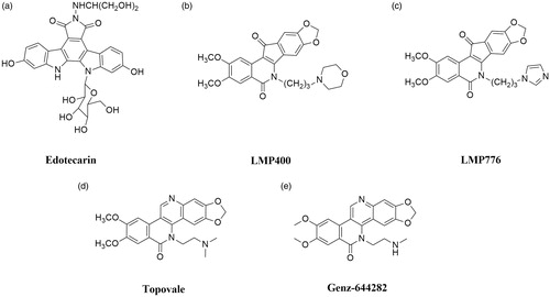
Figure 6. Chemical structure of etoposide.
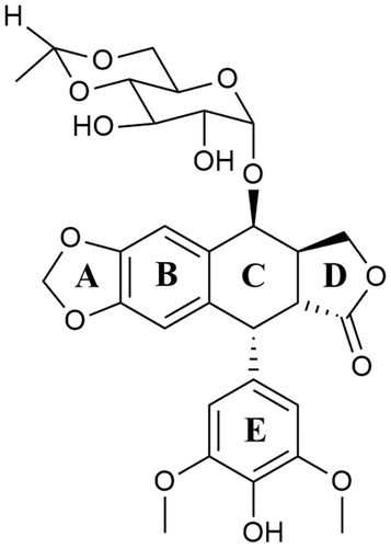
Figure 7. Chemical structures of epipodophyllotoxins.
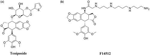
Figure 8. Chemical structures of doxorubicin.
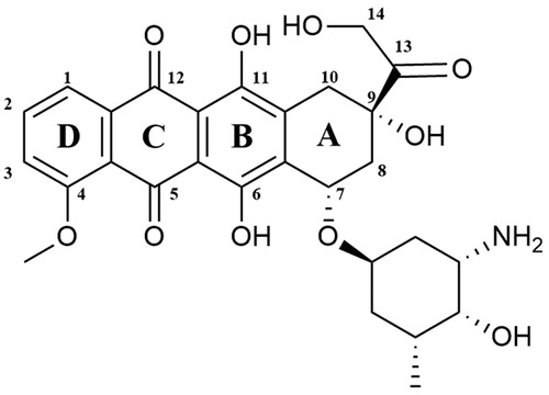
Figure 9. Chemical structure of anthracyclines.
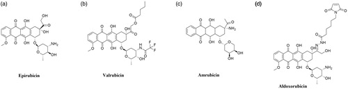
Figure 10. Chemical structure of mitoxantrone.
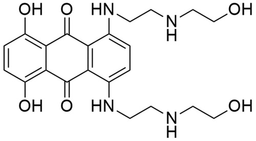
Figure 11. Chemical structure of amsacrine.
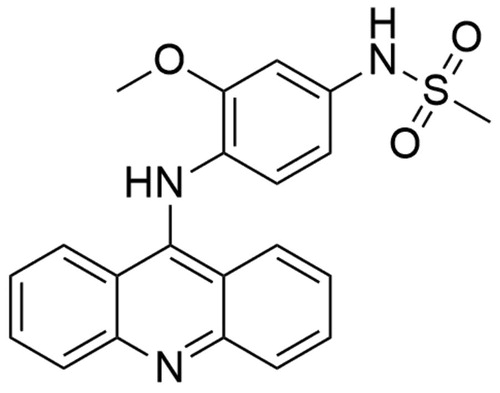
Figure 12. Chemical structure of vosaroxin.
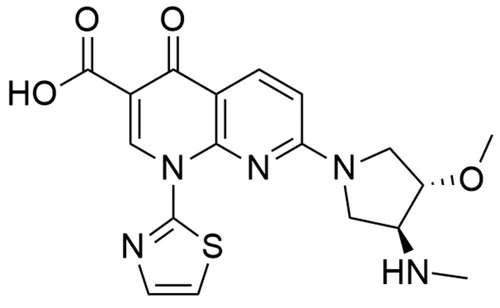
Figure 13. Chemical structure of dexrazoxane.
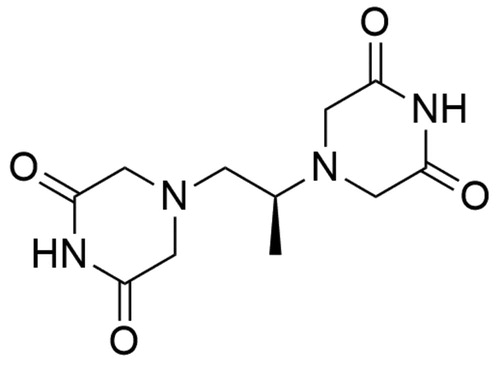
Figure 14. Chemical structure of merbarone.