
Figures & data
Figure 1. Schematic representation of zinc-binding CAI general structure in the CA binding site.
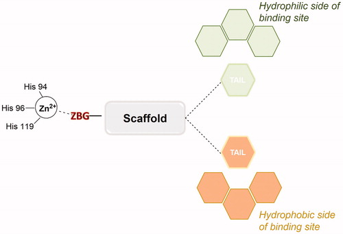
Figure 2. Chemical structures of Acetazolamide (AZA) 1, Brinzolamide 2 and Dorzolamide 3.
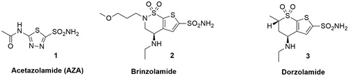
Figure 3. General structures of Topiramate analogues and anomeric sulfonamides, anomeric sulfamides and 6-sulfamoyl carbohydrates.
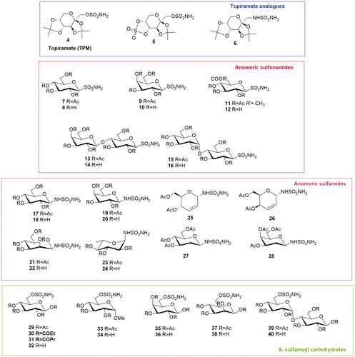
Figure 4. General structures of Topiramate thioureido-derivatives and anomeric sulfamates.
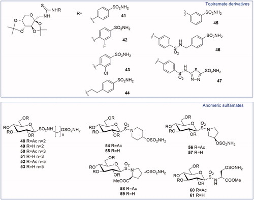
Figure 5. Interactions at the binding site in the structure of CA IX/57 complex (A), CA II/57 complex (B) and CA IX/55 complex (C) Citation29. A) CA IX mimic (cyan) and 57 (magenta) (PDB ID: 4R5A). B) CA II (grey) and 57 (green) (PDB ID: 4R59). C) Overlay of the two conformations of 55 (purple and orange) with CA IX mimic. (PDB ID: 4R5B).
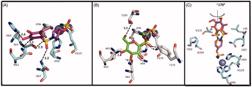
Figure 6. General structures of S-glycosyl sulfonamides and sulfenamides and thioureido-glycoconjugates.

Figure 7. General structures of amido phenyl sulfonamide glycoconjugates.
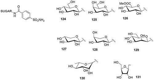
Figure 8. General structures of anomeric 1,4-disubstituted triazole sulfonamide glycoconjugates.
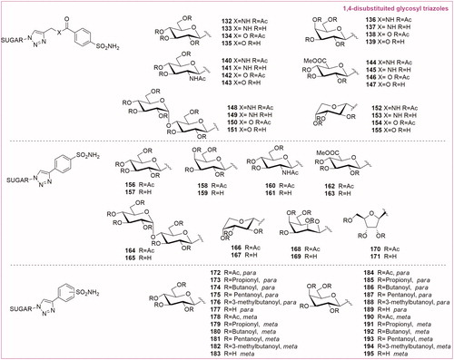
Figure 9. General structures of O-Glycosyl and the S-glycosyl 1,4-disubstituted triazoles, 1,4-disubstituted triazole saccharin-glycoconjugates and 1,5-disubstituted glycosyl triazoles.
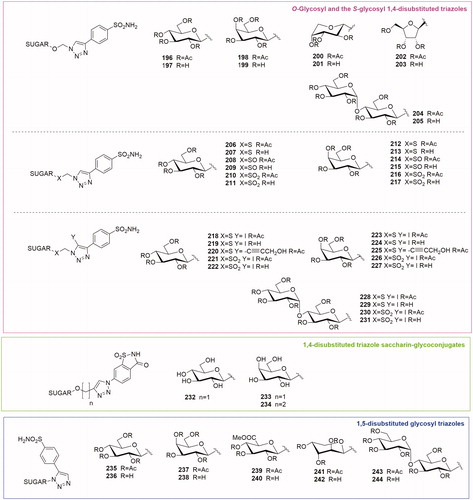
Figure 10. General structure of coumarin glycosylated CA inhibitors.
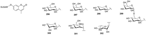
Figure 11. General structure of C-glycosyl CA inhibitors.
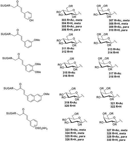
Figure 12. General structure of dual-tail CA inhibitor glycoconjugates.
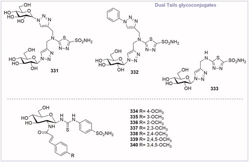
Figure 13. (A) Interactions at the binding site in the structure of CA II/332(magenta) complex (PDB ID: 4CQ0). (B) Surface representation of CA II/332 complex. The hydrophobic half of CA II is red and the hydrophilic half is blue to highlight the different interactionsCitation67.
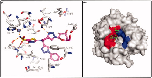
Table 1. Chemical structure, CA IX inhibitory activity, CA I/ CA IX selectivity profile of the best compound for each class of CA glycosidic inhibitors
Table 2. Chemical structure, CA IX inhibitory activity, CA I/ CA IX selectivity profile of the best compound for each class of CA glycoconjugated inhibitors