
Figures & data
Table 1. IC50 values of the aqueous (AE), ethanolic (EE) and methanolic (ME) extracts from W. filifera collected in the areas of Sousse (S) and Gabès (G) against α-glucosidase and α-amylase.
Figure 1. Inhibition of α-glucosidase enzyme. Lineweaver–Burk plots analysis of EEG (a) and MEG (b).
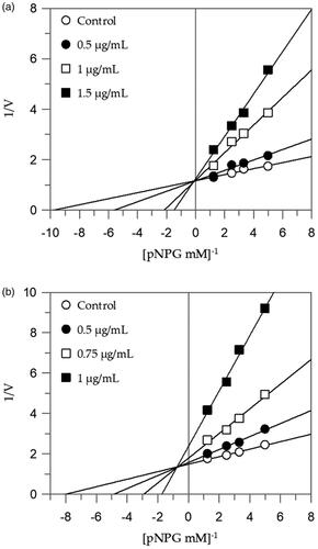
Table 2. Enzyme kinetics parameters following α-glucosidase and α-amylase with different W. filifera seed extracts.
Figure 2. Inhibition of α-amylase enzyme. Lineweaver–Burk plots analysis of EES (a) and MES (b).
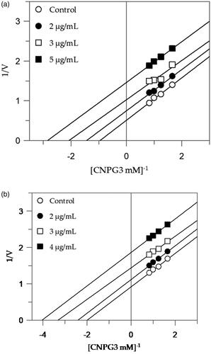
Figure 3. Thioflavin T fluorescence emission plot corresponding to β-sheet formation of IAPP in the presence of W. filifera methanolic seeds extract from Sousse.
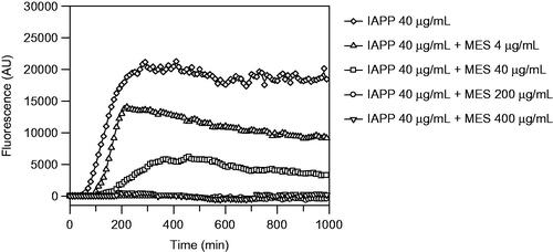
Figure 4. Congo red and Electron microscopy analyses of the material recovered after ThT analysis. In (a), amyloid exhibiting green birefringence after Congo red staining and (b) long unbranched amyloid fibrils are present in solution of IAPP 40 µg/mL. In (c), no Congophilc material can be detected and in (d) an amorphous material is present in solution containing IAPP 40 µg/mL with MES 200 µg/mL. Samples in a and c are stained with Congo red and samples in b and d are negatively contrasted with 2% Uranyl acetate in 50% ethanol.
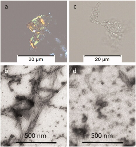
Table 3. Binding energies of against IAPP, α-amylase and α-glucosidase.
Table 4. Physicochemical properties of bioactive compounds in the investigated extracts.