
Figures & data
Figure 1. Active site view of A. hCA II (pdb 1RAY) and B. NgCA (pdb 1KOP) in adduct with the azide anion N3-. Amino acid residues of NgCA are renumbered according to the corresponding residues from hCA II. The zinc ion, represented as a grey sphere, is coordinated by three His residues, that are His94, His96 and His119, and the azide anion. Residues constituting the α-helix portion 128–139 are coloured magenta in hCA II, while being absent in NgCA.
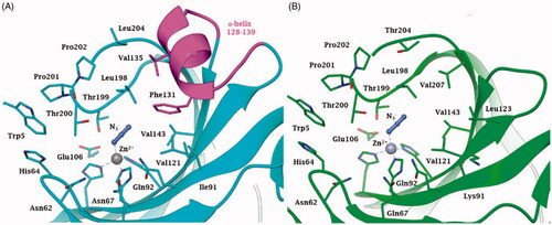
Table 1. Inhibition constants (KIs) of anion inhibitors against hCA I, II and NgCA by a stopped flow CO2 hydration assayCitation17.