Figures & data
Figure 1. (A, B) Effect of dasatinib treatment on serum levels of the liver enzymes; alanine transaminase (ALT) and aspartate transaminase (AST) in mice with thioacetamide-induced liver fibrosis. The data are presented as mean ± SEM (n = 6). aSignificant difference from the control group; bsignificant difference from liver fibrosis-inducted group (at p ˂ .05)
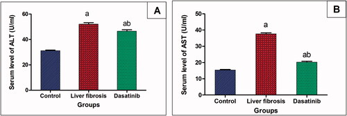
Figure 2. Effect of dasatinib treatment on relative expression of miRNA-378-3p, Wnt-10a and β-catenin in the liver tissue of mice with thioacetamide-induced liver fibrosis. The data are presented as mean ± SEM (n = 6). aSignificant difference from the control group; bsignificant difference from liver fibrosis-inducted group (at p ˂ .05).
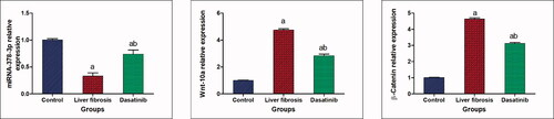
Figure 3. (A–E) Effect of dasatinib treatment on relative expression of miRNA-17-5p, smad-7, smad-3, collagen a1 and Wnt inhibitory factor-1 (WIF-1) in liver tissue of mice with thioacetamide-induced liver fibrosis. The data are presented as mean ± SEM (n = 6). aSignificant difference from the control group; bsignificant difference from liver fibrosis inducted-group (at p ˂ .05).
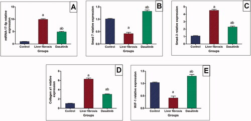
Figure 4. Immunostaining of α-smooth muscle actin (α-SMA) and tumour necrosis factor-α (TNF-α) in the liver tissue of mice with thioacetamide-induced liver fibrosis (H&E × 40). (A) α-SMA/control group, (B) α-SMA/liver fibrosis group, (C) α-SMA/dasatinib-treated group, (E) TNF-α/control group, (F) TNF-α/liver fibrosis group, (G) TNF-α/dasatinib-treated group, (D, H) represent a comparative quantification of the immunohistochemical expression for α-SMA and TNF-α in hepatic tissue of mice from all groups: The severity of the immunoactivity is depending on the intensity and distribution of the brown colour. aRepresents a significant difference from the normal control group, ba significant difference from liver fibrosis inducted-group (at p ˂ .05).
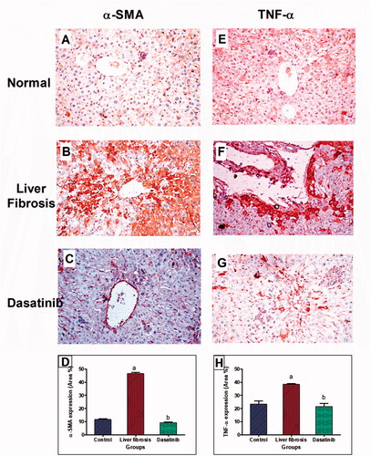
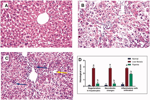
Figure 5. Effect of dasatinib treatment on the histopathological alterations in the liver tissue in mice with thioacetamide-induced liver fibrosis (H&E × 16): (A) control group, (B) liver fibrosis group, (C) dasatinib-treated group, (D) scoring of the histological observations in the hepatic tissue from all groups. Data are presented as mean ± SEM of 6 random non-overlapping fields/section. aSignificant difference from the control group, bsignificant difference from liver fibrosis inducted-group (at p ˂ .05).