Figures & data
Figure 1. Nrf2 activators from natural products or their derivatives and caffeoylisocitric acid.
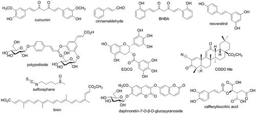
Figure 2. Effects of caffeoylisocitric acid on the survival of mesangiacl cells. (A) Viability of mesangial cells cultured in normal medium. (B) Viability of mesangial cells cultured in medium containging high glucose. (C) BrdU incorporation assay. n = 3, ##P < 0.01 vs control group, *P < 0.05 and **P < 0.01 vs high glucose group.
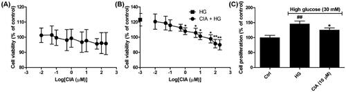
Figure 3. Effects of caffeoylisocitric acid on the activation of Nrf2 in mesangial cells under high glucose. (A) Immunofluorescence staining for Nrf2. (B) Western blot analysis for the expression of nuclear and total Nrf2. (C)-(D) Densitometric analysis for the expression of nuclear and total Nrf2. (E) Relative ARE-luciferase activity. (F)-(I) ELISA assays for the levels of HO-1, NQO1, GST A1 and A2. n = 3, #P < 0.05 and ##P < 0.01 vs control group, ***P < 0.001 vs high glucose group.
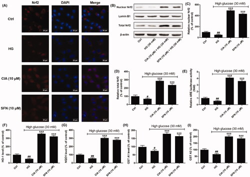
Figure 4. Effects of caffeoylisocitric acid on the oxidative stress induced by high glucose in mesangial cells. (A) Intracellular ROS levels. (B) MDA content. (C)-(D) SOD and CAT activity. n = 3, #P < 0.05 and ###P < 0.001 vs control group, ***P < 0.001 vs high glucose group.
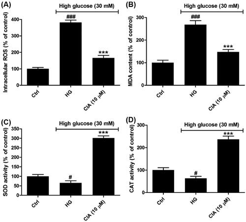
Figure 5. Effects of caffeoylisocitric acid on the inflammation induced by high glucose in mesangial cells. (A) Relative NK-κB-RE luciferase activity. (B)-(D) Relative levels of pro-inflammatory cytokines including TNF-α, IL-1β and IL-6. n = 3, ###P < 0.001 vs control group, **P < 0.01 and ***P < 0.001 vs high glucose group.
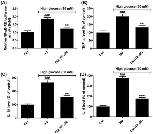
Figure 6. Effects of caffeoylisocitric acid on the accumulation of ECM induced by high glucose in mesangial cells. (A)-(B) Secretion of TGF-β1 and CTGF in mesangial cells. (C)–(E) Synthesis of ECM proteins including collagein IV, fibronectin and laminin. n = 3, ###P < 0.001 vs control group, ***P < 0.001 vs high glucose group.
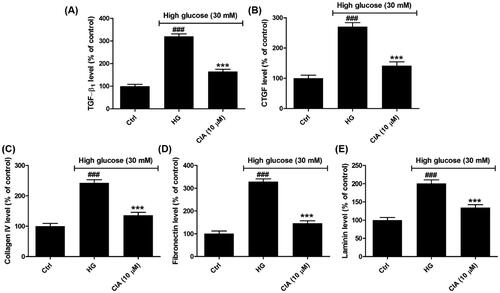
Figure 7. Effects of caffeoylisocitric acid on MAPK signalling pathway in mesangial cells under high glucose. (A)-(C) Levels of p-ERK1/2, p-JNK and p-p38 MAPK in mesangial cells. n = 3, ###P < 0.001 vs control group, **P < 0.01 and ***P < 0.001 vs high glucose group.
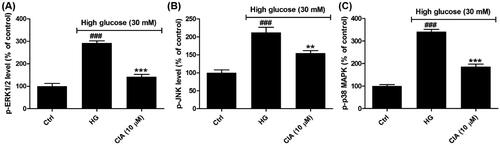
Figure 8. Activation of Nrf2 is involved in the effects of caffeoylisocitric acid in mesangial cells under high glucose. (A) Western blot analysis for the Nrf2 knockdown. (B) Densitometric analysis for the expression of Nrf2. (C)-(J) Intracellular ROS and MDA as well as extracellular collagen IV, fibronectin, laminin, TNF-α, IL-1β and IL-6 in mesangial cells transfected with Nrf2-siRNA or NC-siRNA exposed to caffeoylisocitric acid. n = 3, ###P < 0.001 vs control group, **P < 0.01 and ***P < 0.001 vs high glucose group.
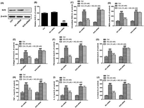
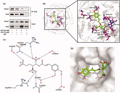
Figure 9. Interaction between Keap1 and caffeoylisocitric acid to activate Nrf2. (A) Co-immunoprecipitation assay for Nrf2. (B)-(D) Molecular docking for Kelch domain and caffeoylisocitric acid.