
Figures & data
Figure 1. Representative anti-angiogenic agents binding to tubulin colchicine binding site.
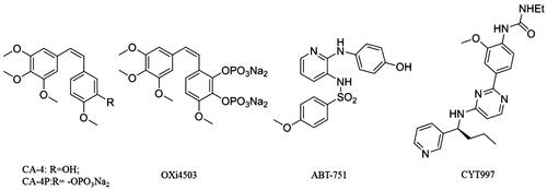
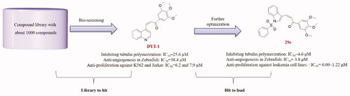
Figure 2. Identification of hit compound (DYT-1) from our chemical library and further optimizations to discover lead compound (29e).
Scheme 1. Reagents and conditions: (i) EtOH, 40% NaOH, 0 °C, 30 min; room temperature, 4 h.

Scheme 2. Reagents and conditions: (i) EtOH, 40% NaOH, 0 °C, 30 min; room temperature, 4 h; (ii) NaH, different haloalkanes, THF, 0 °C to room temperature.
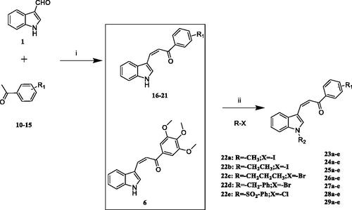
Table 1. Tubulin inhibitory activities and antiangiogenic activities in Zebrafish of chalcone derivatives with variation of the LHS
Table 2. Tubulin inhibitory activities and antiangiogenic activities in Zebrafish of chalcone derivatives with variation of both LHS and RHS.
Table 3. IC50 Values of 29e and CA-4 against various leukaemia cell lines
Figure 3. Compound 29e induced apoptosis K562 cells. (A) Apoptosis ratio detection by flow cytometry assays for 48 h. (B) The quantitative analysis of apoptotic rate at early and advanced stages of K562 cells. (C) Western blot analysis of the apoptosis related proteins. (D) The quantitative analysis of the protein levels. The data was presented as the mean ± SD of three independent tests.
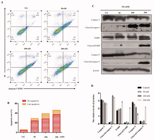
Figure 4. Compound 29e bind to the colchicine site of tubulin and inhibit the microtubule polymerisation. (A) 2 D model of the interaction between compound 29e and the amino acid residues of tubulin colchicine site. (B) 3 D model of the binding position of compound 29e. (C) RMSD tendency of two systems (29e, colchicine) versus time in the 60 ns MD simulation. (D) Effect of compound 29e on tubulin binding of [3H] colchicine. (E) Compound 29e affected microtubule assembly in vitro. (F) The effects of 29e on the organisation of cellular microtubule network of K562 cells.
![Figure 4. Compound 29e bind to the colchicine site of tubulin and inhibit the microtubule polymerisation. (A) 2 D model of the interaction between compound 29e and the amino acid residues of tubulin colchicine site. (B) 3 D model of the binding position of compound 29e. (C) RMSD tendency of two systems (29e, colchicine) versus time in the 60 ns MD simulation. (D) Effect of compound 29e on tubulin binding of [3H] colchicine. (E) Compound 29e affected microtubule assembly in vitro. (F) The effects of 29e on the organisation of cellular microtubule network of K562 cells.](/cms/asset/5c47a6a2-70c6-4a33-b8b7-b7154c3bd29a/ienz_a_2032688_f0004_c.jpg)
Figure 5. Compound 29e showed antivascular activity in vitro. (A) The wound-healing assay was used to evaluate the migration of HUVEC cells, and images were captured at 0 h and 48 h after treatments with 29e. (B) The invasion suppressing effects of 29e against HUVECs cells by Transwell assay. (C) Typical images depicting tubule formation of HUVEC cells by treatments with 29e for 6 h. (D) Quantitative analysis of the migration ability of HUVEC tubule formation.
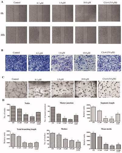
Figure 6. Anti-angiogenic effect of 29e evaluated by zebrafish embryos assay. (A)Inhibitory effect of 29e on the angiogenesis of transgenic zebrafish. (B) Histogram showed the numbers of zebrafish ISVs per field under confocal microscopy.
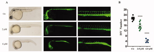
Figure 7. Inhibitory effects of 29e on the proliferation and metastasis of K562 cells in zebrafish xenograft models. CMTPX labelled K562 cells (red) were microinjected into zebrafish embryos, and indicated concentration of 29e were added. White solid arrows indicate disseminated cells.
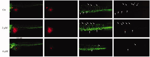
Table 4. Physicochemical parameters and metabolic stability of compound 29e