Figures & data
Figure 1. The essential pharmacophoric features of erlotinib as an EGFR inhibitor occupying three pockets in the ATP binding site based on ReferenceCitation22.
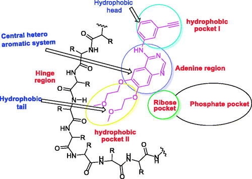
Figure 2. Some reported EGFR-TK inhibitors and their basic pharmacophoric features.
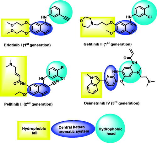
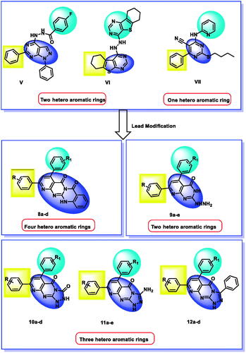
Figure 3. Synthesis of new EGFR inhibitors strategy.
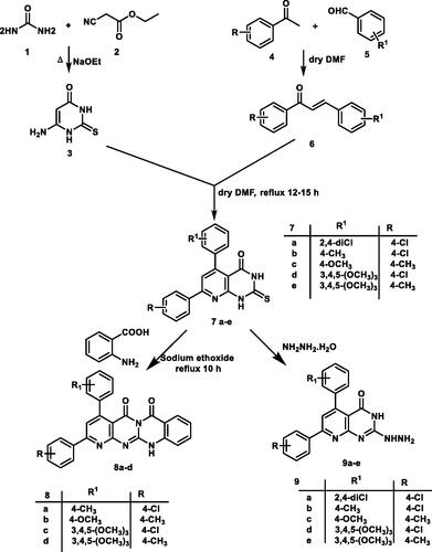
Scheme 1. General procedure for the synthesis of the target compound 7a–e, 8a–d, and 9a–e.
Scheme 2. General procedure for the synthesis of the target compound 10a–d, 11a–e and 12a–d.
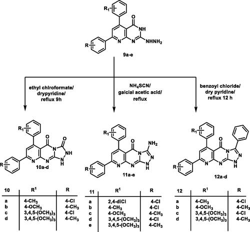
Table 1. Percentage of growth inhibition activity of compounds 7a, 8a–d, 9a, 10a–e against A549, PC-3, HCT-116 and MCF-7 at a concentration of 100 μM.
Figure 4. SAR according to modifiable moieties in the target compounds.
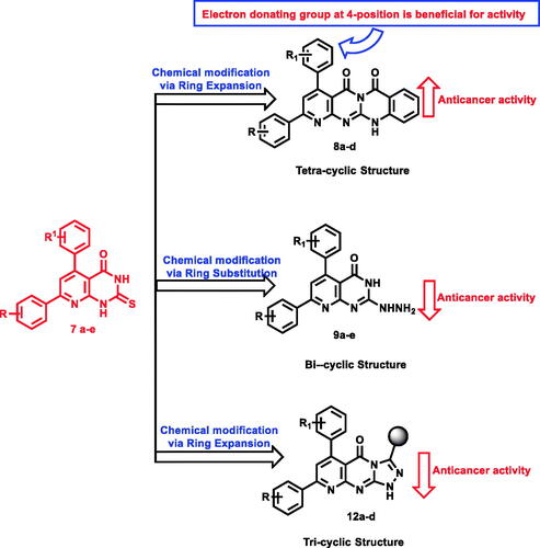
Table 2. IC50 values of compounds 8a, 8b, 8d, 9a and 12b against A-549, PC-3, HCT-116 and MCF-7.
Table 3. In vitro enzymatic inhibitory activities against EGFRL858R and EGFR790M.
Figure 5. PC3distribution upon treatment with compound 8a.
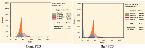
Figure 6. Apoptosis and necrosis percent induced by compound 8a.
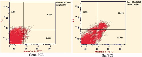
Table 4. Effect of compound 8a on active caspase-3 in PC-3 cells after 24 h treatment.
Table 5. The docking binding free energies of the synthesised compounds against EGFRWT and EGFRT790M.
Figure 7. (A and B) 3D and 2D superimposition of the docked ligand (erlotinib; pink) and the original ligand (green) with RMSD value of 0.88 Å.
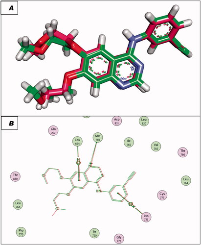
Figure 8. (A and B) 3D and 2D superimposition of the docked ligand of mutant EGFR (TAK-285; Pink) and the original ligand (green) with RMSD value of 1.06 Å.
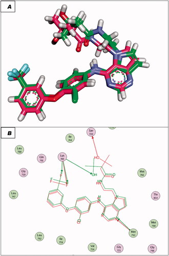
Figure 9. Erlotinib docked into the active site of EGFRWT.
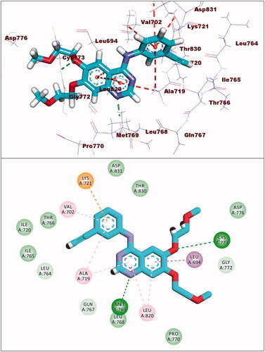
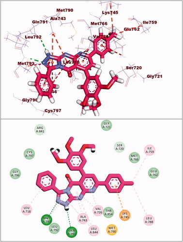
Figure 10. Compound 8a docked into the active site of EGFRWT.
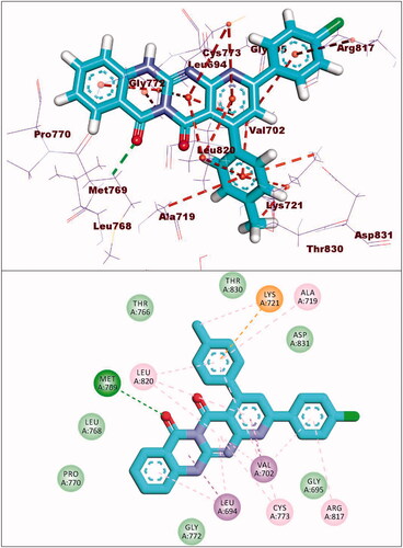
Figure 11. Compound 8b docked into the active site of EGFRWT.
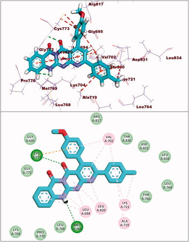
Figure 12. Compound 8d docked into the active site of EGFRWT.
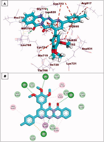
Figure 13. Compound 9a docked into the active site of EGFRWT.
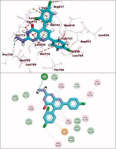
Figure 14. Co-crystallised ligand (TAK-285) docked into the active site of EGFRT790M.
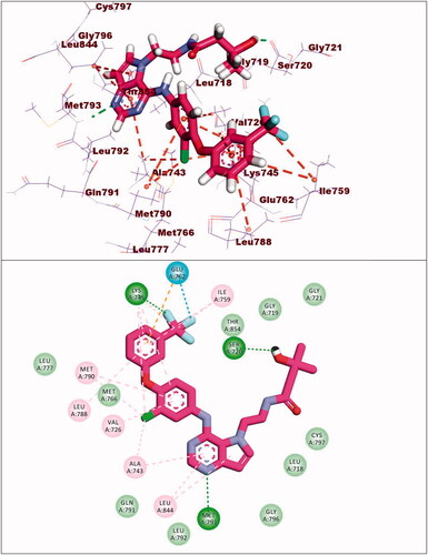
Figure 15. Binding of compound 8c with EGFRT790M.
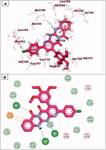
Figure 16. Binding of compound 9a with EGFRT790M.
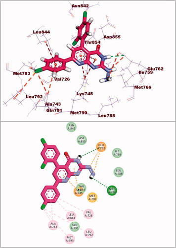
Figure 17. Binding of compound 10d with EGFRT790M.
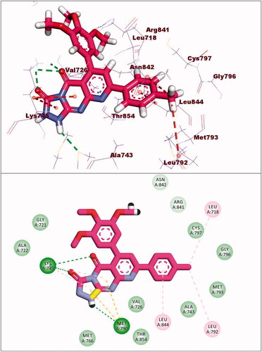
Figure 18. Binding of compound 12d with EGFRT790M.