
Figures & data
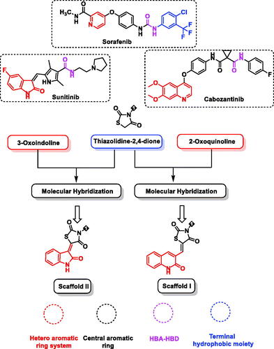
Figure 1. Design of target compounds based on FDA-approved VEGFR-2 inhibitors and molecular hybridisation strategy.
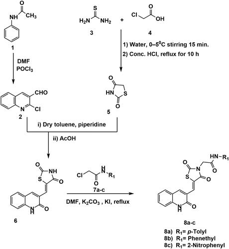
Scheme 1. Synthesis of compounds 8a–c.
Scheme 2. Chemical synthesis of compounds 12a and b.
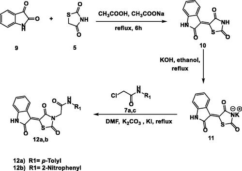
Table 1. In vitro anti-proliferative activities of 8a–c and 12a,b against Caco-2, HepG2, and MDA-MB-231 cell lines.
Table 2. IC50 values of the tested compounds against VEGFR-2 and Vero cell line and their selectivity index (SI) against different cancer cell lines.
Figure 2. Selectivity indices of the synthesised compounds.
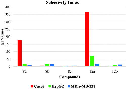
Figure 3. Effect of compound 12a on cells migration and healingefficacy of Caco-2 cells.
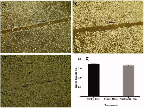
Figure 4. Relative gene expression levels of 4 different genes (BCL2, BCLXL, Survivin, and TGF) in Caco-2 cell line treated with 12a using RT-qPCR.
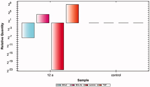
Table 3. Docking binding free energies (ΔG) of the synthesised candidates with VEGFR-2 enzyme.
Figure 5. 3D and 2D binding mode of sorafenib into VEGFR-2 active site.
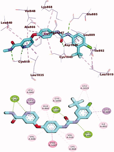
Figure 6. 3D and 2D binding mode of 8a with the active site of VEGFR-2.
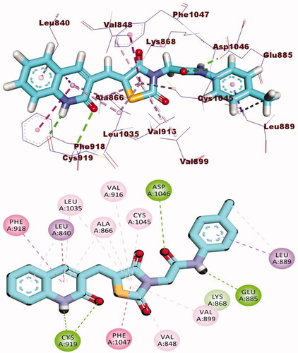
Figure 7. 3D and 2D binding mode of 12a with the active site of VEGFR-2.
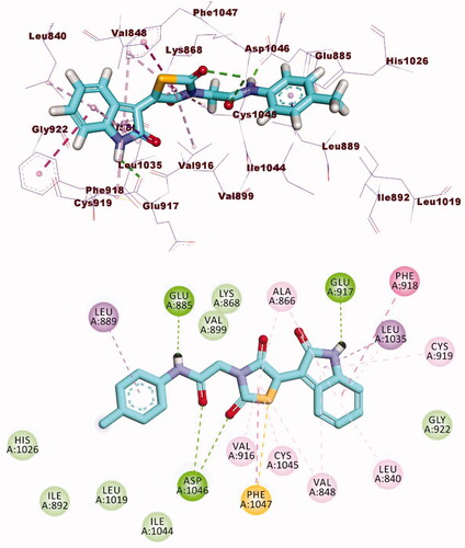
Figure 8. M D simulations experiment: (A) RMSD values of compound VEGFR-2-compound 12a complex before and after binding, (B) RMSF of VEGFR-2-compound 12a complex, (C) Rg of VEGFR-2-compound 12a complex, D) SASA of VEGFR-2-compound 12a complex, E) H- bonding between VEGFR-2-compound 12a complex.
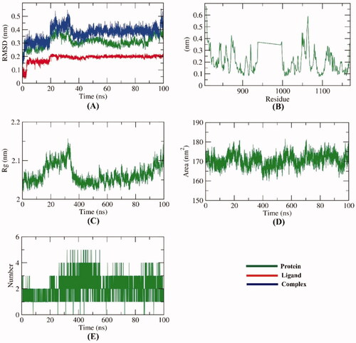
Figure 9. MM-PBSA analysis.
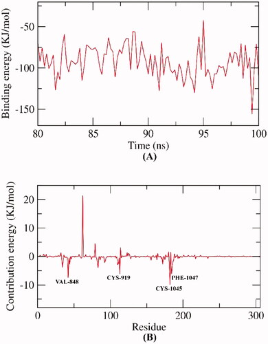
Figure 10. In silico predicted ADMET parameters for the synthesised compounds and references.
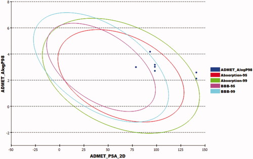
Table 4. ADMET parameters for the synthesised compounds and reference molecules.
Table 5. In silico toxicity of the synthesised compounds and reference molecules.