Figures & data
Figure 1. Chemical structures of resveratrol (Res) and pterostilbene (Ptb) and their positive effects in the brain. On the right side the structure of Ptb factionalised on phenolic ring with benzofurazan 1 (yellow) and with rhodamine B-isothiocyanate 2 (pink), respectively.

Scheme 1. (i) K2CO3, anhydrous DMF, 50 °C, 8 h, then r.t., 36 h. (ii) CF3COOH, DCM, r.t., 12 h, Et2O • HCl, 0 °C. (iii) LiOH, NDB H2O/THF, r.t., 12 h. (iv) NaOH 0.1 M. (v) DMF, Rhodamine-B Itc, r.t, 48 h.
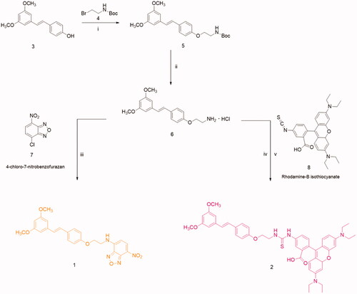
Table 1. Maximum absorption wavelengths and ε values of NBD, Res, 1 and 2.
Table 2. Excitation wavelength, emission wavelength and emission intensity (a.u.) values of NBD, Res, 1 and 2.
Figure 2. Absorption and emission spectra collected on 1 and 2.
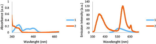
Figure 3. Carbon K-edge spectrum (a), nitrogen K-edge spectrum (b), and oxygens K-edge spectrum (c) of samples 1 and 2.
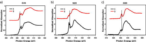
Figure 4. FTIR spectra of samples 2 (a), 1 (b) and of 7-nitrobenzofurazan (c).
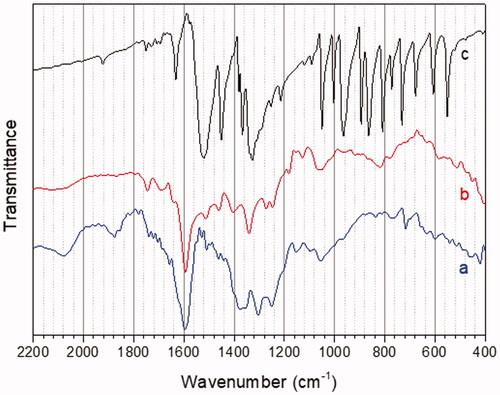
Figure 5. Analyses of cellular DNA content obtained from propidium iodide assay (PI) in SH-SY5Y neuroblastoma (left) and MCF-7 breast cancer (right) cells. (a) Cells were treated for 48 h with either vehicle (ethanol/DMEM, 1/10) or resveratrol (Res, 1 µM) or with different Pterostilbene (Ptb) or its fluorescent derivatives (1 and 2) concentrations. Data are means ± SD of height (SH-SY5Y) or five (MCF-7) different experiments. (b) Typical Western blot of five different experiments of PARP-1 (uncleaved 116 kDa, cleaved 89 kDa) in cells treated as in (a).
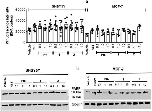
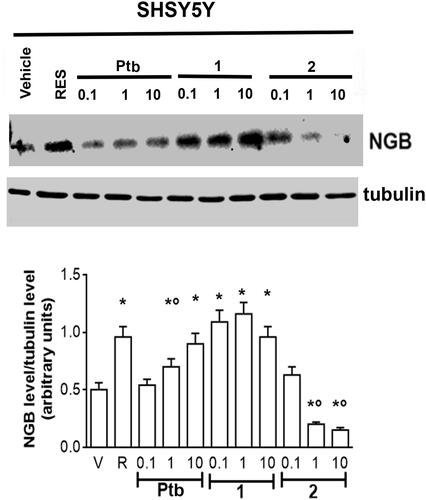
Figure 6. Effect of pterostilbene (Ptb) and its fluorescent derivatives (1 and 2) on neuroglobin (NGB) levels in neuronal-derived cells. Western blot (upper panel) and densitometric analyses (bottom panel) of NGB levels in SH-SY5Y cells. Cells were treated for 24 h with resveratrol (R, 1 µM) or with different compound concentrations. The amount of NGB was normalised by comparison with α-tubulin levels. Data are the mean ± SD of five different experiments. p < 0.001 was determined with ANOVA followed by Bonferroni test with respect to the vehicle (*) or to R-treated samples (°).