1,831
Views
2
CrossRef citations to date
0
Altmetric
Research Paper

Combining nano-differential scanning fluorimetry and microscale thermophoresis to investigate VDAC1 interaction with small molecules

, , , , &
Article: 2121821 | Received 01 Aug 2022, Accepted 01 Sep 2022, Published online: 17 Jan 2023

Figures & data

Figure 1. Structure of the open state of hVDAC1 in LDAO micelles (PDBID:2JK4)Citation15. Left: side (membrane) view. Right: top (cytoplasm) view. The barrel pore is coloured in blue and the alpha helix in yellow.

Figure 1. Structure of the open state of hVDAC1 in LDAO micelles (PDBID:2JK4)Citation15. Left: side (membrane) view. Right: top (cytoplasm) view. The barrel pore is coloured in blue and the alpha helix in yellow.

Table 1. List of selected compounds including experimental techniques used to show direct interaction as well as observed effect(s) on VDAC1 and corresponding references.

Figure 2. Thermal stability of hVDAC1 in LDAO micelles determined by NanoDSF in presence or absence of 5% DMSO. The S-shaped transition of the ratio of tryptophan fluorescence at 350 and 330 nm is displayed in the top figure while corresponding first derivatives are shown in the bottom figure. Inflection points, which correspond to the Tm, are indicated by dashed lines. Tm values were 57.60 ± 0.05 °C and 56.00 ± 0.07 °C in the absence or in the presence of 5% DMSO, respectively. Standard deviations were obtained by performing triplicates of each experiment.

Figure 2. Thermal stability of hVDAC1 in LDAO micelles determined by NanoDSF in presence or absence of 5% DMSO. The S-shaped transition of the ratio of tryptophan fluorescence at 350 and 330 nm is displayed in the top figure while corresponding first derivatives are shown in the bottom figure. Inflection points, which correspond to the Tm, are indicated by dashed lines. Tm values were 57.60 ± 0.05 °C and 56.00 ± 0.07 °C in the absence or in the presence of 5% DMSO, respectively. Standard deviations were obtained by performing triplicates of each experiment.

Figure 3. First derivative of the 350/330 nm ratio obtained from NanoDSF as a function of the temperature. Only compounds displaying a significant Tm shift are shown. (A) Compounds solubilised in buffer. (B) Compounds solubilised in DMSO. Reference Tm, obtained for hVDAC1 (in absence (A) or in presence of 5% DMSO (B)) is indicated.

Figure 3. First derivative of the 350/330 nm ratio obtained from NanoDSF as a function of the temperature. Only compounds displaying a significant Tm shift are shown. (A) Compounds solubilised in buffer. (B) Compounds solubilised in DMSO. Reference Tm, obtained for hVDAC1 (in absence (A) or in presence of 5% DMSO (B)) is indicated.

Figure 4. MST binding check performed on 100 nM hVDAC1 with (green), or without (blue) 250 µM VBIT4.

Figure 4. MST binding check performed on 100 nM hVDAC1 with (green), or without (blue) 250 µM VBIT4.

Figure 5. MST dose-response curve of the eight compounds displaying precise or minimum Kd values (cannabidiol, curcumin, DIDS, emodin, itraconazole, VBIT4, propofol and fluoxetine). Error bars show standard deviation obtained from three replicas of the same measurement.

Figure 5. MST dose-response curve of the eight compounds displaying precise or minimum Kd values (cannabidiol, curcumin, DIDS, emodin, itraconazole, VBIT4, propofol and fluoxetine). Error bars show standard deviation obtained from three replicas of the same measurement.

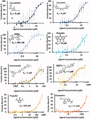
Table 2. Results obtained by NanoDSF and MST including Tm shift, MST binding check and dissociation constant evaluated for each tested compounds and published affinity if available. A thermal shift was considered significant when its variation exceeded ± 0.22 °C. (N.D., not determined, when experiments were not performed due to inconclusive binding check assay; N.A., not available, when experiments were performed but were inconclusive). hVDAC1 = human recombinant VDAC1 and rVDAC1 = VDAC1 extracted from rat.

Supplemental material

Supplemental Material

Download PDF (117.7 KB)