
Figures & data
Figure 1. Chemical structures of synthetic compounds 1a–19a and 1b–19b.Citation30
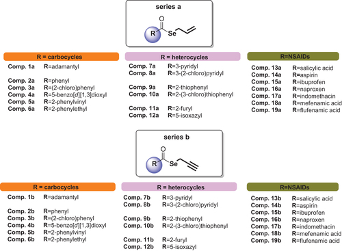
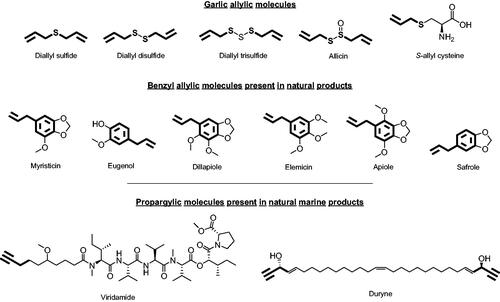
Figure 2. Allylic and propargylic molecules of natural products.Citation30
Scheme 1. Synthesis procedure to yield 1a–19a and 1b–19b derivatives. Reagents and conditions: (i) H2O, 20 min, and room temperature; (ii) THF/H2O, 60 min, and room temperature; (iii) ClCOCOCl, CH2Cl2, 12 h, and room temperature; (iv) BrCH2CH = CH2 or BrCH2C≡CH, THF/H2O, 90 min, and room temperature.Citation30
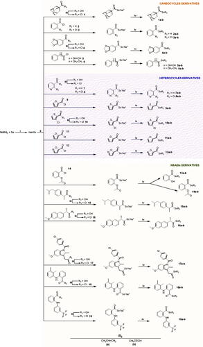
Table 1. Inhibition of hCA I, II, VA, IX, and XII activity for compounds 1a–19a and 1b–19b.
Figure 3. Predicted binding mode of propargyl selenol (violet) and allyl selenol (cyan) within the (A) CA I (yellow), (B) CA II (white), (C) CA IX (blue) and (D) CA XII (green) active site.
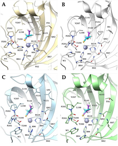
Figure 4. (A) Superposition of (A) X-ray solved structures of phenyl selenol-CA II (red, PDB:6HX5)Citation25 and benzyl selenol-CA II (green, PDB:7QBH)Citation53 complex. (B) Superposition of ligands benzyl selenol (green), propargyl selenol (cyan), and allyl selenol (violet). (C) Superposition of X-ray solved structures of benzyl selenol-CA II (green)Citation53 and predicted binding mode of allyl (violet)/propargyl (cyan) selenol within CA II active site.
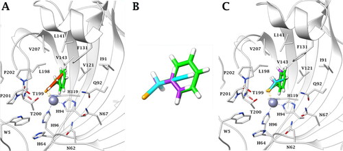
Table 2. Geometric parameters of docking and crystallography data.