Figures & data
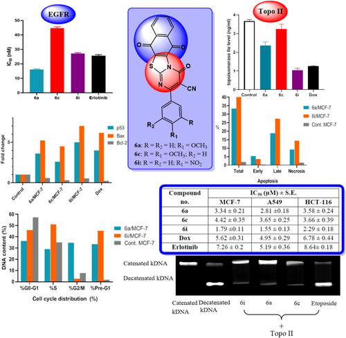
Figure 1. Examples for anticancer agents targeting topoisomerase II.
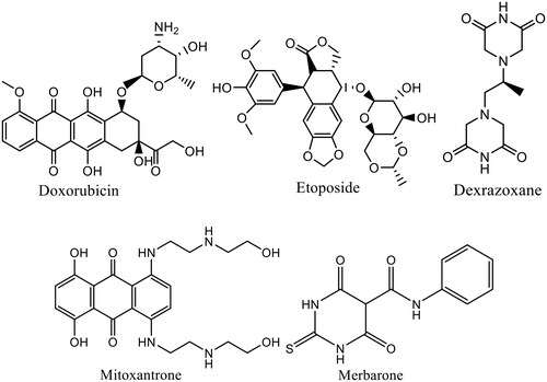
Figure 2. Clinically approved EGFR anticancer drugs.
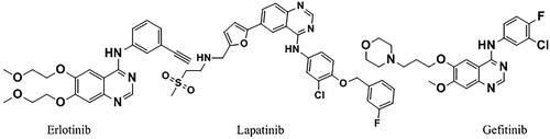
Figure 3. Diagram represents design of novel hybrids based upon thiazolo[3,2-a] pyrimidine and 1,4-naphthoquinone moieties as dual topo II/EGFR inhibitor.
![Figure 3. Diagram represents design of novel hybrids based upon thiazolo[3,2-a] pyrimidine and 1,4-naphthoquinone moieties as dual topo II/EGFR inhibitor.](/cms/asset/8316e3c5-4555-436f-906b-6d26001b0569/ienz_a_2205043_f0003_c.jpg)
Scheme 1. Synthesis of the target hybrids 6a-i. Reagents and conditions: a) K2CO3, C2H5OH, reflux, 2h. b) DMF , rt., 12 h.
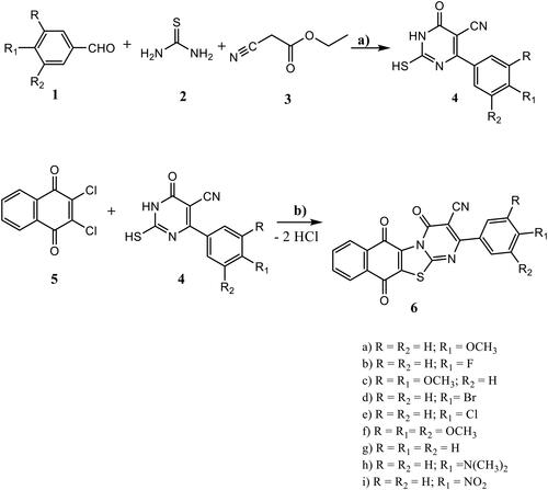
Figure 4. A representative example of 1H NMR and 13C NMR of 6i hybrid.
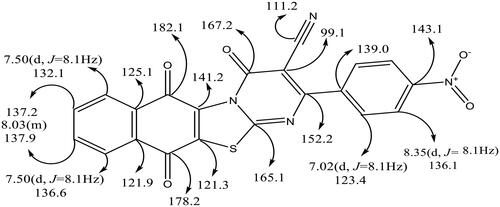
Table 1. Cytotoxicity of the synthesised hybrids (6a-i), dox and erlotinib against MCF-7, A549 and HCT-116 cell lines.
Figure 5. Effect of the tested hybrids (6a, 6c and 6i) and Dox on topoisomerase IIα concentration in MCF-7 cell line.
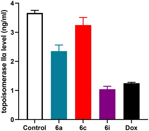
Figure 6. DNA topo II inhibition assay of 6i, 6a and 6c.
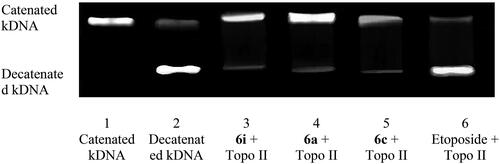
Figure 7. IC50 values of the tested hybrids (6a, 6c and 6i) and erlotinib against EGFR.
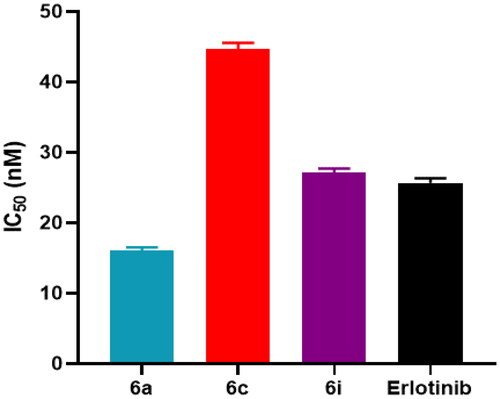
Figure 8. Effects of 6a, 6c and 6i hybrids and Dox on p53, Bax and Bcl-2 levels in MCF-7 cancer cell line.
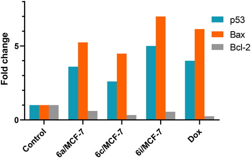
Table 2. Effects of 6a, 6c and 6i hybrids on p53, Bax and Bcl-2 levels in MCF-7 cancer cell line.
Table 3. Effects of 6a, 6c and 6i hybrids on caspase-7 and caspase-9 levels in MCF-7 cancer cell line.
Figure 9. (A) Cell cycle analysis in MCF-7 cell line treated with 6a and 6i hybrids. (B) Cell cycle analysis and apoptosis effect in MCF-7 cell line treated with 6a and 6i hybrids.
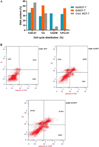
Table 4. Cell cycle analysis of 6a and 6i hybrids in MCF-7 cell line.
Figure 10. (A) Percentage of apoptosis and necrosis for 6a and 6i hybrids in MCF-7 cell line. (B) Flow cytometric analysis of Annexin V-FITC/PI induced by 6a and 6i hybrids in MCF-7 cell line.
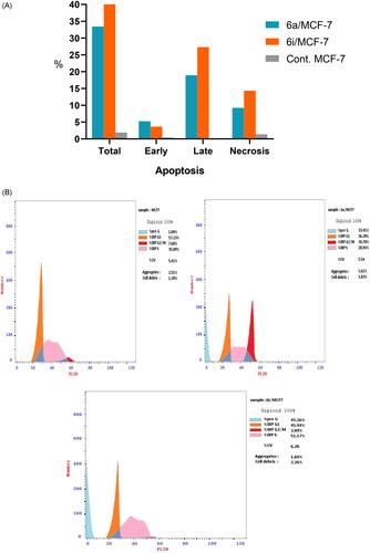
Table 5. Results of apoptotic assay of 6a and 6i hybrids in MCF-7 cell line.
Table 6. Pharmacokinetic prediction of the synthesised hybrids (6a-i) by Molinspiration v2021.03.
Table 7. Bioactivity score of the synthesised hybrids (6a-i).
Table 8. Absorption properties of the synthesised hybrids (6a-i).
Table 9. Distribution properties of the synthesised hybrids (6a-i).
Table 10. Metabolism and excretion properties of the synthesised hybrids (6a-i).
Table 11. Toxicity properties of the synthesised hybrids (6a-i) and Dox.
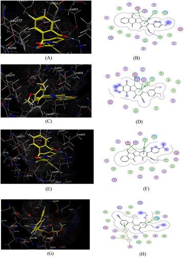
Figure 11. Docking and binding pattern of hybrids 6a (A&B), 6c (C&D), 6i (E&F) and Dox. (G&H) showing interactions with different amino acid residues found in the active site of human topo IIα ATPase (PDB code 1ZXM).
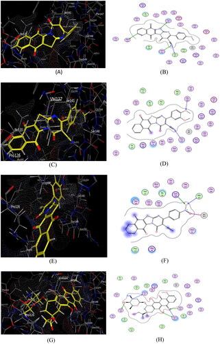
Table 12. Types of binding interactions and energy scores (kcal/mol) for hybrids (6a–i) and Dox at the human topo IIα ATPase/AMP-PNP active site.
Table 13. Types of binding interactions and energy scores (kcal/mol) for hybrids (6a–i) and erlotinib at the EGFR kinase active site.
Figure 12. Docking and binding pattern of compounds 6a (A&B), 6c (C&D), 6i (E&F) and erlotinib (G&H) showing interactions with different amino acid residues found in the ATR-active site of EGFR (PDB code: 1XKK).