
Figures & data
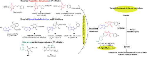
Figure 1. Design of new 5-arylidene-2,4-TZDs-based hybrids as ARIs based on some reported AR inhibitors.
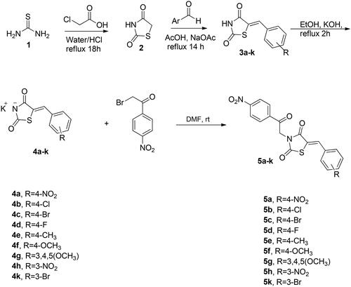
Scheme 1. Synthetic pathway for the target 4-nitro-phenacyl tethered thiazolidine-2,4-dione hybrids 5a–k.
Scheme 2. Synthetic pathway for the target benzothiazole tethered thiazolidine-2,4-dione hybrids 8a–h.
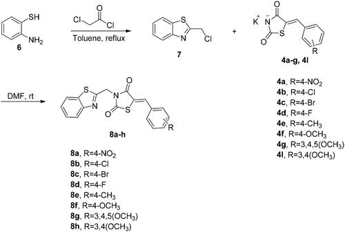
Table 1. IC50 values of the tested compounds 5a–k and 8a–h against human aldose reductase.
Figure 2. Lineweaver–Burk plot showing kinetics of aldose reductase in the absence and presence of different concentrations of 8b.
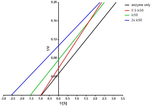
Figure 3. In vivo hypoglycaemic effect of compound 8b in STZ-induced diabetes model after 6 weeks of treatment. Data shown are averages of six independent experiments *p > 0.05 and ****p > 0.0001.
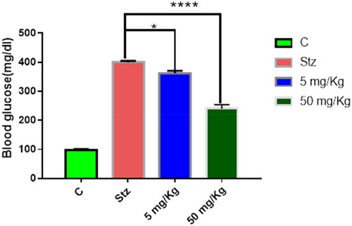
Figure 4. Molecular docking of 8b in the active site of aldose reductase PDB: 3g5e. (A) Compound 8b (green) aligned with the co-crystallised ligand (red). (B) 3D binding interaction of the hybrid 8b with active site residues of aldose reductase. (C) 2D interaction of compound 8b with active site residues of aldose reductase.
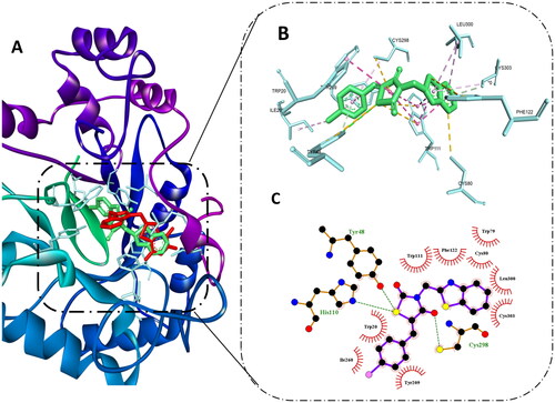
Table 2. Calculated parameters for Lipinski’s rule and Veber’s standards for the hybrids 5a, 5f, 8b, 8c, 8e, and 8g.
Table 3. ADMET profile for the active hybrids 5a, 5f, 8b, 8c, 8e, and 8g.