Figures & data
Figure 1. (a) 33-year-old female subject with baseline BMI of 29. (b) 16-week follow-up: subject’s left flank was treated and correctly identified by both blinded reviewers. Subject reported on the 5-point Likert Scale that she was very satisfied with treatment outcome and that the results were highly visible.
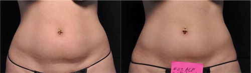
Figure 2. (a) 53-year-old female subject with baseline BMI of 22. (b) 16-week follow-up: subject’s left flank was treated and correctly identified by both blinded reviewers. Subject reported on the 5-point Likert Scale that she was neutral with treatment outcome and that the results were slightly visible.
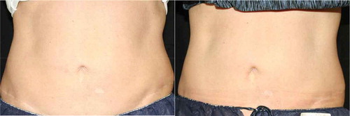
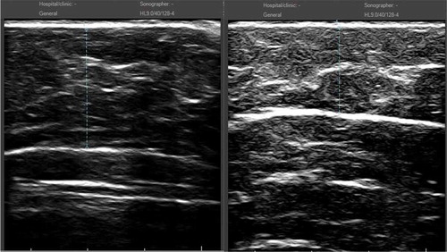
Figure 3. (a) Ultrasound image for 33-year-old female subject () prior to treatment. (b) 16-week follow-up: measurement showed 4.2 mm reduction (from 13.8 mm at baseline to 9.6 mm at follow-up).
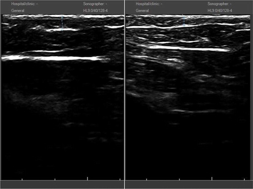
Figure 4. (a) Ultrasound image for 53-year-old female subject () prior to treatment. (b) 16-week follow-up: measurement showed 7.0 mm reduction (from 21.2 mm at baseline to 14.2 mm at follow-up).