
Figures & data
Figure 1. Thirty-two-year-old patient, G2P1, 62 days gestation, no lower abdominal pain or vaginal bleeding. Blood β-HCG 114,384 IU/ml. Ultrasound examination showed an elongated gestational sac of which the lower extremity located at the cesarean section scar (anterior lower segment) with scattered anechoic foci within chorion.
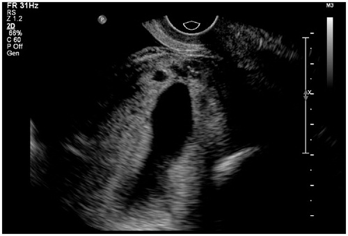
Figure 2. Thirty-seven-year-old patient, G3P1, 52 days gestation. She had suffered from vaginal bleeding for 12 days without lower abdominal pain. Blood β-HCG level 77,198 IU/ml. Ultrasound showed a gestational sac totally located in the lower segment cavity and protruded from uterus contour. Calipers located at the outer edge of gestational sac. CX: cervix; F: uterine cavity fluid.
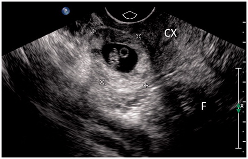
Figure 3. Thirty-nine-year-old patient, G4P1, 75 days gestation. She complained vaginal bleeding for a few days. Blood β-HCG 230 IU/ml. Ultrasound detected a thickened wall gestational mass within lower uterine segment. Multiple anechoic foci of various sizes with hypo-echoic dots could be seen within the thickened wall. The double-head arrow pointed out the thickness measurement of chorion.
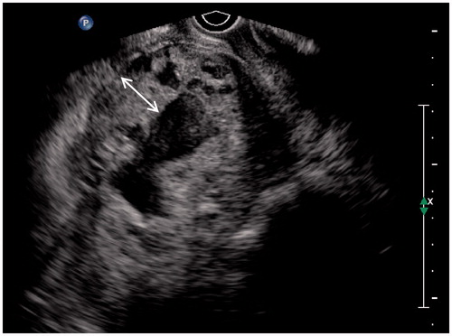
Figure 4. Thirty-six-year-old patient, G4P1, 50 days gestation, vaginal bleeding (+). Blood β-HCG 168,136 IU/ml. Ultrasound showed an elongated gestational sac with lower extremity implanted in the uterine lower segment (between calipers). There was no lacunar-like change of chorion membrane.
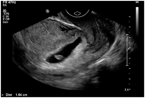
Figure 5. Measurement of minimum thickness of uterine lower segment myometrium: the hypoechoic myometrial layer (double-headed arrow) between the outer edge of the chorion (lower dotted line) and the inner edge of the uterine serosa (upper dotted line) was taken for measurement.
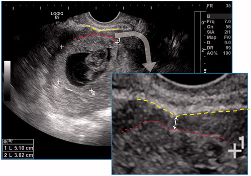
Figure 6. Grading of the blood signal on color Doppler flow imaging (CDFI) of uterine lower segment: (a) CDFI grade 1, very little blood signal; (b) CDFI grade 2, moderate blood signal; (c) CDFI grade 3, rich blood signal (from the same patient of ).
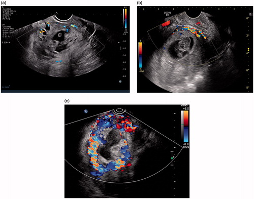
Table 1. Comparison of clinical manifestations and laboratory test of cesarean scar pregnancy (CSP) patients in early pregnancy.
Table 2. Comparison of sonographic features of cesarean scar pregnancy (CSP) patients in early gestation.
Table 3. Comparison of treatment methods and hospitalization of cesarean scar pregnancy (CSP) patients in early gestation termination.