
Figures & data
Figure 1. Measurement of the length and maximum transverse diameters of the fetal dilated small duodenum by ultrasound.
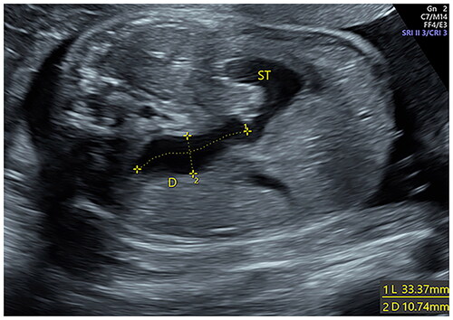
Figure 2. Measurement of the longitudinal dimension of dilated stomach by ultrasound.
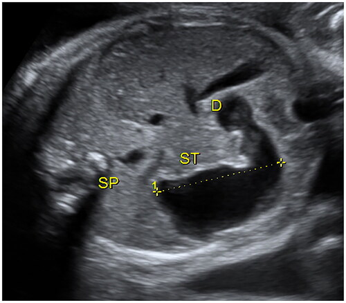
Figure 3. (a) A fetus was diagnosed annular pancreas by postnatal surgery. Prenatal ultrasound shown that the dilated bowel connected with the stomach, and the end of the enlarged intestine was located at the level of right side of spine at 35 weeks. White arrow indicates the stomach; yellow arrow indicates the end of the enlarged intestine; blue arrow indicates the spine. (b) A fetus was diagnosed high jejunal atresia by postnatal surgery. Prenatal ultrasound shown that the enlarged intestine, in a “C” shape, connected with the stomach, and extended from the right side of the abdominal cavity to the left side, and the end of the enlarged intestine significantly exceeded the level of left side of spine at 29 weeks. White Arrow indicates the stomach; yellow arrow indicates the end of the enlarged intestine; blue arrow indicates the spine.
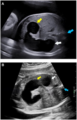
Table 1. Clinical characteristics of patients with CDO and high jejunal obstruction.
Table 2. Prenatal sonographic findings of patients with CDO and high jejunal obstruction.
Figure 4. The ROC curves in the prenatal diagnosis of fetal CDO.
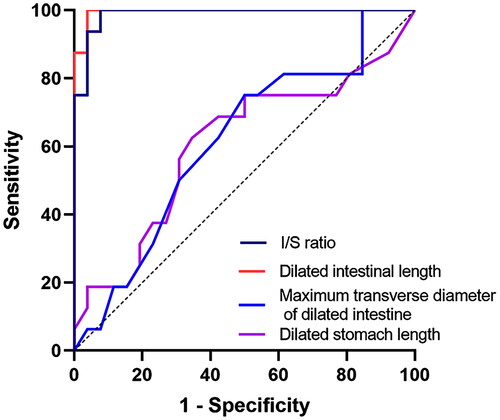
Table 3. Results of receiver operating characteristic curve analysis.
Table 4. Correlation between the location of CDO and ultrasound metrics.