
Figures & data
Figure 1. Intraperitoneal calcification in a prenatal ultrasound scan. AS: ascites; STO: stomach. The bold arrows indicate calcification on the surface of the liver and intestines.
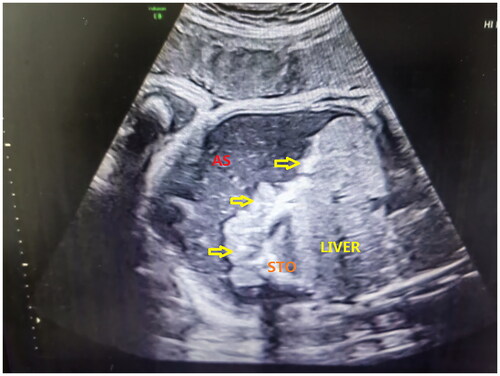
Figure 2. Intestinal dilatation in a prenatal ultrasound scan. RK: right kidney; LK: left kidney; Thick arrows represent calcified, thickened, and dilated intestinal tube walls.
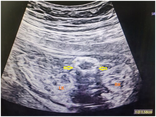
Table 1. Comparison of general data of neonates between the operation group and conservative treatment group (X ± SD).
Table 2. Results of prenatal ultrasonography in the operation group and conservative treatment group.
Table 3. Surgical method in the operation group.
Table 4. The maternal complications in different groups.
Data availability statement
The datasets used and/or analyzed during the current study available from the corresponding author on reasonable request.